
- Title
-
Quality assurance of hematopoietic stem cells by macrophages determines stem cell clonality
- Authors
- Wattrus, S.J., Smith, M.L., Rodrigues, C.P., Hagedorn, E.J., Kim, J.W., Budnik, B., Zon, L.I.
- Source
- Full text @ Science
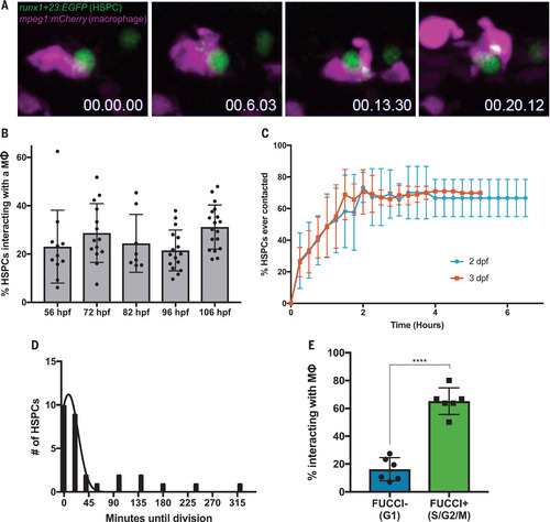
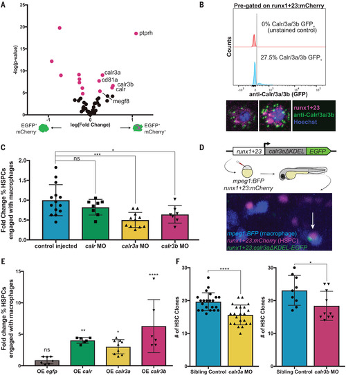
|

|
|

|