
- Title
-
egfl6 expression in the pharyngeal pouch is dispensable for craniofacial development
- Authors
- Jin, S., Na, H., Jeon, H., Park, J., Choe, C.P.
- Source
- Full text @ Animal Cells Syst (Seoul)

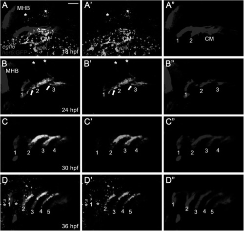
Expression of egfl6 in pouch formation. (A-D) Fluorescence in situ hybridization of egfl6 (green) in conjunction with the GFP immunohistochemistry (red) in wild-type Tg(her5:GFP) animals. (A) At 18 hpf, egfl6 expression is seen in the her5-positive second (2) pouch and the posterior cell mass (CM), with no egfl6 expression seen in the first (1) pouch. egfl6 expression is also seen in the developing hindbrain (asterisks). Note of non-specific green staining in the yolks (dotted line). (B) At 24 hpf, egfl6 is expressed in all three her5-positive pouches (1-3), with new egfl6 expression appearing in the mesoderm between pouches (arrows). egfl6 expression is still seen in the developing hindbrain (asterisks). (C) At 30 hpf, egfl6 expression is only observed in all four her5-positive pouches (1-4), with the egfl6 expression in the mesoderm gone. (D) At 36 hpf, egfl6 is expressed in all pouches, with its expression in the fifth (5) pouch being faint. Also, unidentified tissues adjacent to the first (1) and second (2) pouches express egfl6 (asterisks). Note that the sixth pouch is barely seen at the level of tissues. MHB: midbrain−hindbrain boundary. (A′–D′) Green channel only. (A”–D”) Red channel only. Scale bar: 40 μm. |
Expression of egfl6 in pouch formation. (A-D) Fluorescence in situ hybridization of egfl6 (green) in conjunction with the GFP immunohistochemistry (red) in wild-type Tg(her5:GFP) animals. (A) At 18 hpf, egfl6 expression is seen in the her5-positive second (2) pouch and the posterior cell mass (CM), with no egfl6 expression seen in the first (1) pouch. egfl6 expression is also seen in the developing hindbrain (asterisks). Note of non-specific green staining in the yolks (dotted line). (B) At 24 hpf, egfl6 is expressed in all three her5-positive pouches (1-3), with new egfl6 expression appearing in the mesoderm between pouches (arrows). egfl6 expression is still seen in the developing hindbrain (asterisks). (C) At 30 hpf, egfl6 expression is only observed in all four her5-positive pouches (1-4), with the egfl6 expression in the mesoderm gone. (D) At 36 hpf, egfl6 is expressed in all pouches, with its expression in the fifth (5) pouch being faint. Also, unidentified tissues adjacent to the first (1) and second (2) pouches express egfl6 (asterisks). Note that the sixth pouch is barely seen at the level of tissues. MHB: midbrain−hindbrain boundary. (A′–D′) Green channel only. (A”–D”) Red channel only. Scale bar: 40 μm. |

Generation of loss-of-function mutations in egfl6 gene. (A) Structure of egfl6 gene. egfl6 gene consists of 13 exons bearing sequences for the protein-coding region (black box) and the 5′ and 3′ untranslated regions (open box). The gRNA target site in the second exon is marked in yellow. (B) Mutant alleles of egfl6 gene. The in/del mutation of each mutant allele is shown in the multiple sequence alignments, with the gRNA target and the PAM sites being marked in red and blue, respectively in the wild-typeTU egfl6 sequence. The electrophoretograms show the lesion in each egfl6 mutant allele that is underlined in the multiple sequence alignments. The start loss mutation in egfl6 gene in the wild-type TU strain is highlighted in yellow. (C) Schematic of the Egfl6 protein encoded by the wild-type and mutant alleles. The conserved five domains marked in the wild-type Egfl6 protein are missing in all mutant Egfl6 proteins. |

Normal craniofacial development in egfl6 mutants. (A–D) In both wild-type (A, n = 92) and egfl6 mutant (B, n = 31) embryos at 34 hpf, immunohistochemistry for Alcama (green) shows five pouches (1–5). Reduction of egfl6 with a MO in wild-type (C, n = 64) or egfl6 mutant (D, n = 24) animals display normal five pouches. Sensory ganglia are indicated with asterisks. (E-H) Fluorescent in situ hybridization for rag1 (green) at 4 dpf. In both wild-type (C, n = 74) and egfl6 mutant (D, n = 29) zebrafish, rag1 is expressed normally in the thymus. Reduction of egfl6 in wild-type (G, n = 61) or egfl6 mutant (H, n = 17) animals shows normal thymus. (I-L) Ventral whole-mount views of dissected facial cartilages at 5 dpf. Both wild-type (E, n = 105) and egfl6 mutant (F, n = 37) zebrafish invariantly form a triangled shape of hyomandibular (HM) and five ceratobranchial (CB) cartilages on each side. egfl6-MO-injected animals in wild-type (K, n = 88) or egfl6 mutant (H, n = 15) animals have normal facial cartilages, including the HM and CBs. Scale bar: 40 μm. |

Expression of egfl7 in the pharyngeal region. (A, B) Fluorescence in situ hybridization of egfl7 (green) in conjunction with the GFP immunohistochemistry (red) in wild-type animals at 30 hpf. (A) egfl7 is expressed segmentally in small patches (arrows) adjacent to her5-positive pouches (1-4). (B) egfl7 expressing small patches (arrows) are located at the ventral tip of sox10-positive pharyngeal arches (PA2-4) but rarely overlapped with PAs. (C-E) Alcama immunohistochemistry (green) labels five pouches (1-5) in wild-type (n = 92), egfl7-MO (n = 80), and egfl7-MO-injected egfl6 mutant (n = 21) embryos at 34 hpf. Sensory ganglia are indicated with asterisks. (F-H) At 4 dpf, rag1 expression (green) in the thymus is normal in wild-type (n = 74), egfl7-MO (n = 76), and egfl7-MO-injected egfl6 mutant (n = 14) zebrafish. (I-K) Facial cartilages, including the HM and CBs, are normal in wild-type (n = 105), egfl7-MO (n = 84), and egfl7-MO-injected egfl6 mutant (n = 19) animals at 5 dpf. Scale bar: 40 μm. |