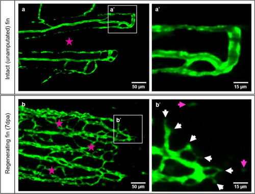
Vascular organization of intact caudal fin vs. early regeneration. Organization of tissue compartments in the intact caudal fin (a) versus the regenerating fin (b), at 7dpa. a/a’ display smooth and hierarchically well-organized blood vessels (green - reporter transgenic zebrafish line) in which intraray segments do not contain any vessels (asterisk) and capillary sprouts are not detectible (a’). (b/b’) display dense, well-interconnected capillary meshwork. The intraray region (purple asterisks) contains bridging vessels and at the expanding vascular front multiple sprouts (b’, white arrows) and migrating - endothelial cells (purple arrows). Images are acquired by fluorescent reflected light microscope.

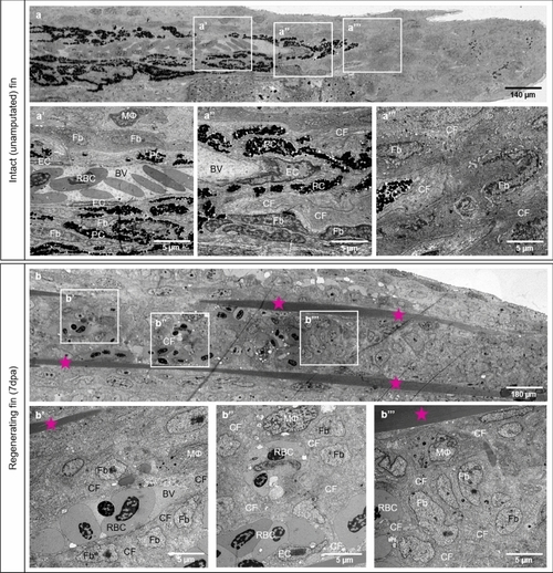
Differences in tissue compartments between the intact (unamputated) caudal fin and fin during early regeneration. Structural differences between intact caudal fin (a) and early regeneration at 7dpa (b) are demonstrated by transmission electron microscopy. a-a’’ display perfused and mature blood vessel with quiescent and well-differentiated endothelial cells (ECs), pigment cells (PC) and ECM containing robust collagen fibers (CF), fibroblasts (Fb) and very seldom solitary macrophage (MΦ). In the regenerating fin (7dpa), robust collagen fibers (CF) are arranged into two densely packed stripes tracing the future ray bones (b, asterisk). Between the latter, new formed, immature blood vessel (BV) containing red blood cells (RBC) are enclosed by scaffold of CF tracing the direction of blood vessel expansion (b’). The CFs are surrounded tightly by Fb and MΦ (b’). At the vascular front, next to new formed capillaries containing ECs, clusters or single RBC are detectible (b’’). In the blastema, multiple Fb proliferate displaying classical mitotic figures; ECM contains CF and MΦ (b’’’). PC are absent during the regenerative phase. Left side - zebrafish trunk, right side - zebrafish tail, images acquired by electron microscope.

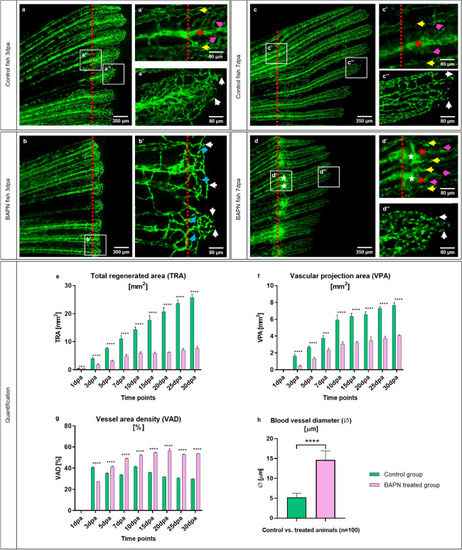
In vivo effects of the collagen cross-linking inhibitor, BAPN, on caudal fin regeneration and neoangiogenesis. Vascular alteration during normal fin regeneration (a, c) versus BAPN-treated group (b, d) at 3dpa and 7dpa is documented (green - reporter transgenic zebrafish line). Vessels in the regenerating region of the controls (a, a’ and c, c’, red dotted lines indicate the amputation) appear hierarchically well organized, building a vascular tree with arteries (red arrow), veins (yellow arrows) and connecting capillaries (purple arrows). Capillaries at the vascular front are representing multiple sprouts (a’’, c’’, white arrows). Shorter regeneration area and respectively modest vascularization in BAPN-treated animals (b); hierarchical organization in supplying and draining vessels as well as capillaries is less pronounced. Many capillary connections are very thin and not perfused (b’, blue arrows); multiple sprouts are visible at the vascular front (b’, white arrows). At 7dpa the regenerating region, respectively the vascular plexus is smaller in comparison to the controls. Balloon-like enlarged blood vessels (detectible as a green sphere) are visible at the site of amputation (d, d’, asterisks). Those angioma-like structures are connected to the arteries (d’, red arrow) and in some cases to the veins (d’, yellow arrows). Further distal tiny connecting capillaries are indicated by purple arrows (d’): vessel network is disorganized and contains higher number of ECs as indicated by the dense nuclear appearance. Images are acquired with a fluorescent reflected light microscope. Quantification of tissue regeneration and vascularization after inhibition was assessed by three variables: Total regenerated area (TRA = regenerated fin in mm2; e), vascular projection area (VPA = vessels within regenerated part in mm2; f) and vessel area density (VAD = vessels density within the regenerated part in %; g) during a period of 30 days in control group (green) versus BAPN-treated group (purple). n = 5. h) Quantification of lumen dimensions (in μm) between control and BAPN-treated animals. Blood vessel diameter in BAPN-treated animals increases by 70%; n = 100.

Collagen cross-linking inhibitor BAPN causes structural changes in the collagen content and blood vessel formation. a, b, c, d demonstrated reconstructed overview of the fish ray containing bones, cartilage, blood vessels and blastema. Amputation plane by the control group 3dpa (a’, a’’, a’’’) and 7dpa (c’, c’’, c’’’) versus BAPN-treated group at 3dpa (b’, b’’, b’’’) and 7dpa (d’, d’’, d’’’) is shown. aa’’ display perfused, normal blood vessel with well-differentiated endothelial cells (EC), red blood cells (RBC), solitary pigment cells (PC), bones, cartilage (Ca) and compact, dense ECM containing collagen cross-linked fibers (CF). CF are a natural compartment of the cartilage (Ca) during the early regenerative phase (a’); they are also a major component of ECM and located along blood vessel making long sleeves (a’’, arrows) or enclosing the fibroblasts (Fb) and tissue macrophages (MΦ). Higher magnification revealed classical collagen cross-linked fibers (a’’’). In the BAPN-treated fish, at the 3dpa, the intraray regions are enlarged containing dilated blood vessels with impacted RBCs and extravasal RBCs in front of the latter, i.e. bleeding (b, b’). The EC appear seldom (b’’, b’’’, arrows), cover only partially the capillaries and the RBCs are in a direct contact with the amorphous ECM (b’’’). Classical cross-linked CFs are not detectible; instead, Collagen fibrils (Cfl) appear as amorphous material (b’’, b’’’). c/c’ display amputation plane by the control group at 7dpa. Perfused and normal blood vessel is covered by well-differentiated ECs. ECM is compact and dense containing PC, Fb and collagen cross-linked fibers. Collagen is present within the cartilage (Ca) and along blood vessel forming long sleeves (c’’, c’’’, arrows). d/d’ display amputation plane by the BAPN-treated group at 7dpa in which balloon-like enlarged blood vessels (BV) containing densely packed RBCs are documented. Those angioma-like structures are perfused and contains partially ECs (d’’, green arrows). MΦ and Fb are embedded in ECM with loose and amorphous appearance due to the absent classical cross-linked CF; instead of it Cfl are widely presented (d’’, d’’’), left side - zebrafish trunk, right side - zebrafish tail, images acquired by electron microscope.

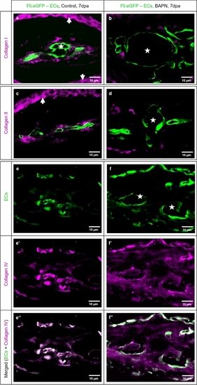
Collagen cross-linking inhibitor BAPN affects collagen I, II and IV accumulation, appearance and distribution. Double immunofluorescence staining revealed the relationship between the new formed blood vessels and collagen I, II, IV distribution in control (left panel: a, c, e, e’, e’’) vs. BAPN-treated animals (right panel b, d, f, f’, f’’) at 7dpa. ECs are labeled green and collagen I, II, IV purple. In the control group, the capillaries have classical appearance and collagen I (a) and collagen II (c) are surrounding the blood vessels as well defined, almost continuous layer. Additionally, collagen I and II stripes are strongly present in the dermal region (arrows). BAPN-treated fish represent dramatically enlarged blood vessels with balloon-like appearance (asterisks). The collagen I (b) and collagen II (d), when present, appear sparse, as small solitary fragments randomly distributed and not associated with the vasculature and dermal region. In the control group collagen IV demonstrated classical co-localization with the EC as a major component of the basement membrane (e, e’, e’’). In the BAPN-treated animals, collagen IV appears disintegrated and diffuse, without co-localization with the ECs (f, f’, f’’). e’’ and f’’ display merged image of ECs and collagen IV corresponding to e, e’ and f, f’ respectively. Images are acquired by confocal microscope.

Ablation of collagen 1α2 producing cells by NFP impair caudal fin regeneration and vascular development. In the control animals, at 3dpa and 7dpa, classical tissue regeneration pattern and blood vessel morphology are documented (a, c). Multiple active cells producing collagen 1α2 (in red) are present adjacent to the amputation line (a’, a’’, c’). Those cells are not detectible in NFP inhibited animals (b, b’, d, d’). Regenerative area and vascular plexus appear shorter and underdeveloped in the NFP-treated group at both, 3dpa and 7dpa. Arrows indicated sprouts. Green - ECs, red dotted line - amputation plane, images acquired by fluorescent reflected light microscope. a’’ part of a’ at higher magnification. Quantification of the regeneration and vascularization after the elimination of the collagen 1α2 producing cells has been performed by three variables: Total regenerated area (TRA = regenerated fin in mm2; (e), vascular projection area (VPA = vessels growth within regenerated fin in mm2; (f) and vessel area density (VAD = vessels density within the regenerated fin in %; g) during the period of 30 days in control group (blue) versus NFP-treated group (red). n = 5.

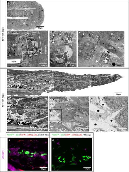
NFP treatment causes collagen deficiency and impairment of caudal fin regeneration. Electron microscopy performed on longitudinal sections revealed severe structural alteration after NFP treatment. 3dpa (a) and 7dpa (b) represented overview of the fish ray containing bones, RBCs deposition and blastema. a’a’’’ display clusters and single RBCs that are in direct contact with surrounding macrophages MΦ, and areas of atypical, amorphous ECM. The latter contains disintegrated cells and cell debris (asterisk) as well apoptotic cells (a’’’, arrow). In contrast to the controls and BAPN treatment, granulocytes (Gr) and MФ are common features. Classical collagen fibers are not detectible. Amputation plane at 7dpa containing perfused vessels is displayed in b; the endothelial coverage is partially interrupted; the EC are activated containing round nuclei and multiple intravascular protrusions. ECM contains less collagen fibers (CF), those do not build classical bundles. The intercellular space is loose, amorphous with irregular distribution representing electron microscopically “empty areas” (b’’’, asterisks). Immunofluorescence staining on transversal sections at the amputation site in control animal (c) and NFP-treated animal (d) at 3dpa are documented. Blood vessels (ECs) are labeled in green, active collagen 1α2 producing cells in red and collagen I purple (c). c demonstrates intact blood vessels, active collagen 1α2 producing cells, and accumulation of collagen I in the vicinity of the blood vessels. d displays blood vessels, very tiny scattered fragments of collagen I and absence of active collagen 1α2 producing cells.

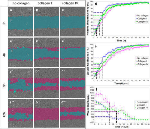
In vitro ECs migration on collagen I and collagen IV substrates. The blue region denotes the scratch wound mask over time (0 h, 4 h, 8 h, and 12 h). ECs migrate into the wound area on non-coated (a), collagen I-coated (b) and collagen IV-coated wells (c). The red area depicts the wound closure by EC expansion over time. The most effective and fast EC migration is observed with the collagen I substrate, followed by collagen IV and lastly, the least efficient, non-coated wells at 0, 4, 8, and 12 h (ac’’’; d, e, and f). Relative wound density (d), wound confluence (e) and wound width (f) has been quantified. Cells in the wound area are the most confluent in collagen I-coated wells, followed by collagen IV and non-coated wells (e). Blue dashed line - collagen I, green dashed line - collagen IV, purple dashed line - no collagen, black dashed line - time points (4 h, 8 h, 12 h).

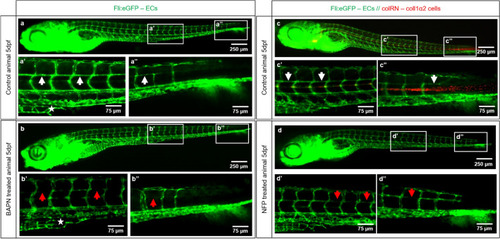
Collagen cross-linking inhibition and reduced collagen 1α2 impair blood vessel formation. Vascular development (ECs - green) in the zebrafish embryo at 5dpf in a control animals (a, a’, a’’ and c, c’, c’’) versus BAPN-treated (b, b’, b’’) and NFP-treated (d, d’, d’’) animals is documented. a’/a’’ shows normal caudal plexus formation with horizontal capillaries connecting the intersegmental vessels (ISV; a’, a’’, white arrow) and subintestinal vein (SIV) (a’, asterisk). The BAPN-treated animals show an underdeveloped plexus with absent SIV (b’, asterisk) and an impairment in the formation of capillaries connecting ISV (b’, b’’, red arrows). cc’’ show normal caudal plexus formation with capillaries connecting ISV (white arrows) and multiple active cells producing collagen 1α2 (in red). Upon NFP treatment, active collagen 1α2 producing cells are absent (dd’’) and the formation of horizontal capillaries connecting the ISV is impaired (d’, d’’, red arrows). ISV - intersegmental vessels, SIV - subintestinal vein; images are acquired by fluorescent reflected light microscopy.

Acknowledgments
This image is the copyrighted work of the attributed author or publisher, and ZFIN has permission only to display this image to its users. Additional permissions should be obtained from the applicable author or publisher of the image. Full text @ Sci. Rep.