FIGURE SUMMARY
Title

Prediction of Sensitivity and Efficacy of Clinical Chemotherapy Using Larval Zebrafish Patient-Derived Xenografts of Gastric Cancer

Authors
Zhai, J., Wu, J., Wang, Y., Fan, R., Xie, G., Wu, F., He, Y., Qian, S., Tan, A., Yao, X., He, M., Shen, L.
Source
Full text @ Front Cell Dev Biol

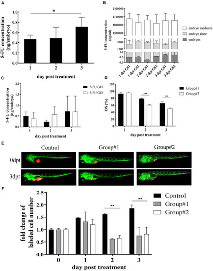
Optimization of 5-FU dosing in zebrafish. (A) The internal concentration of 5-FU in embryos engrafted with SGC7901 cells was increased significantly after treatment with 5-FU (5 mM) in embryo medium, and the average internal concentration of 5-FU was 0.47, 0.49, and 0.71 ng per embryo on 1, 2, and 3 dpt, respectively (*P < 0.05). (B) Treatment for the embryos without xenograft showed that the internal concentration of 5-FU in embryos increased remarkably in both groups (P < 0.05), but the internal concentration was lower than that in embryo mediums and in the embryo rinse, indicating 5-FU enriched in embryos was a tiny fraction of administration [G#1 (group #1), daily refreshment of drug-containing medium; G#2 (group #2), continuous drug-containing medium without refreshment]. (C) zCDX study with SGC7901 cells showed that the internal concentration of 5-FU did not show difference between these two groups on each dpt (P > 0.05). However, the overall survival (OS) of embryos increased in group #1 (***P < 0.0001) (D). (E, F) These two administrations of 5-FU inhibited the tumor cell proliferation in the zCDX with SGC7901 cells (dyed with red fluorescence using DiL) significantly (**P < 0.01), and there was no difference in cell growth inhibition between group #1 and group #2 (P > 0.05).

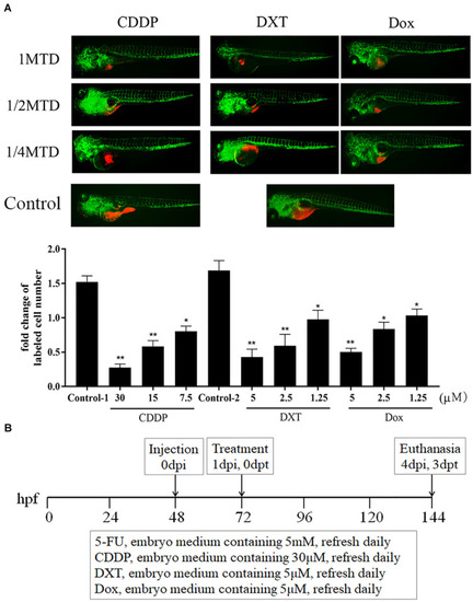
CDDP, DXT, and Dox dosing in zebrafish. (A) zCDXs engrafted SGC7901 cells (dyed with red fluorescence using DiL) were treated with CDDP, DXT, or Dox at their 1/4, 1/2, and 1 MTD, and they inhibited cell proliferation in a dose-dependent manner (control-1, embryo medium for CDDP assay; control-2, embryo medium containing 0.1% DMSO for DXT or Dox assay) (*P < 0.05, **P < 0.01 vs. control-1 or control-2). (B) A protocol for preclinical chemosensitivity test in the zebrafish larvae cancer model was developed.

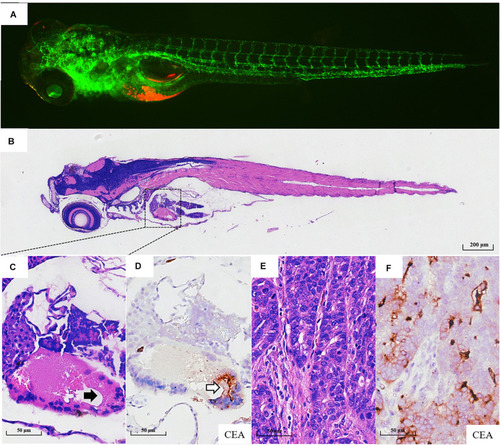
Pathological assays of the engrafted zebrafish for patient #43. (A) The confocal image and (B) the H & E image of the engrafted zebrafish (4 dpi). (C) The enlarged yolk sac showing the tumor xenograft (black arrow indicating engrafted tumor cells), which did not develop to the adenoid structure as that in the patients (E). (D) CEA expression in tumor xenograft (blank arrow). (E) The H & E image of the primary tumor. (F) CEA expression in primary tumor tissue.

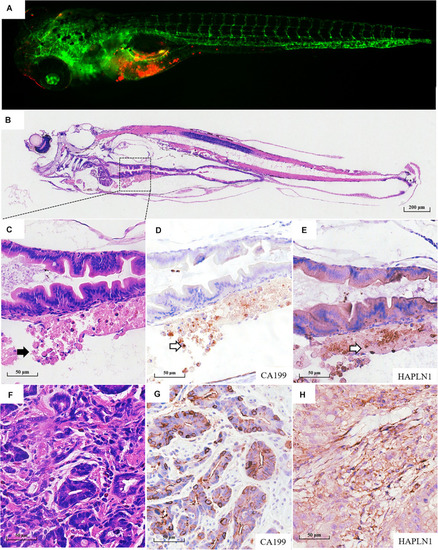
Pathological assays of the engrafted zebrafish for patient #55. (A) The confocal image and (B) the H & E image of the engrafted zebrafish (4 dpi). (C) The enlarged yolk sac showing the tumor xenograft (black arrow indicating engrafted tumor cells), which did not develop to the adenoid structure as that in the patients (F). (D) CA199 expression in tumor xenograft (blank arrow) and (G) CA199 expression in primary tumor tissue. (E) HAPLN1 expression in tumor xenograft (blank arrow) and (H) HAPLN1 expression in primary tumor tissue.

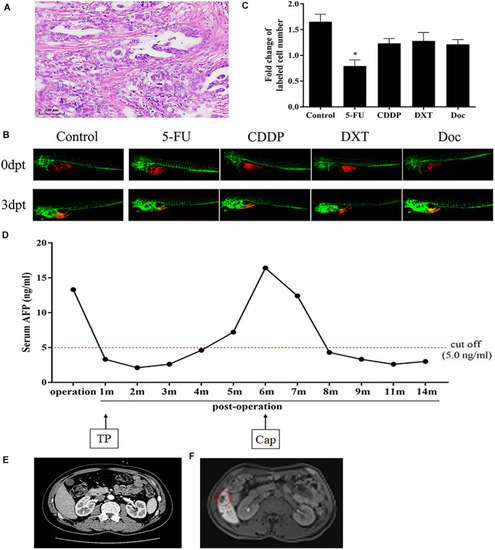
Chemosensitive profiling in zPDXs of patient #20 and its clinical relevance. (A) Patient #20 was diagnosed as diffuse-type gastric adenocarcinoma. (B,C) His zPDXs study showed that 5-FU was the only sensitive drug (*P < 0.05). (D) The patient had elevated preoperative serum AFP level (13.30 ng/ml), and his serum AFP level went down to normal range after radical surgery. He initially received postoperative TP (oxaliplatin/paclitaxel) therapy, but his serum AFP increased to 16.4 ng/ml at postoperative 6 months. (E) He received CT scan at postoperative 1 month as baseline and no abnormality was found. (F) However, his MR monitoring showed one metastatic foci (red ring) in the right lobe of his liver at postoperative 6 months. Thus, he was considered to relapse. The regimen was adjusted to capecitabine (Cap, a prodrug of 5-FU). Two cycles later, his serum AFP level returned to the normal range, and stayed for more than 5 months.

Acknowledgments
This image is the copyrighted work of the attributed author or publisher, and ZFIN has permission only to display this image to its users. Additional permissions should be obtained from the applicable author or publisher of the image. Full text @ Front Cell Dev Biol