FIGURE SUMMARY
Title

A comprehensive structural, lectin and immunohistochemical characterization of the zebrafish olfactory system

Authors
Villamayor, P.R., Arana, Á.J., Coppel, C., Ortiz-Leal, I., Torres, M.V., Sanchez-Quinteiro, P., Sánchez, L.
Source
Full text @ Sci. Rep.

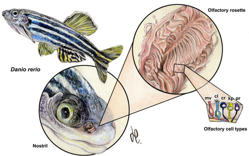
Macroscopic anatomy of the zebrafish olfactory organ. The olfactory rosette occupies in the head an anterodorsal position. The nostrils lie slightly rostral to the eyes and close to the mouth. Five neurosensorial olfactory cell types (OSNs) have been described: microvillous (mv); ciliated (cl); crypt (cr); kappe (kp); and pear (pr) OSNs. Drawing by Helena Reino Piñeiro published under a CC BY open access license.

Microscopic anatomy of the zebrafish olfactory system. (A) Low power sagittal section of the anterior zebrafish stained with Gallego’s trichrome. (B) Higher magnification of the inset in (A), showing the olfactory rosette and the olfactory bulb (OB). (C) Histological section of the olfactory sensory epithelium stained with Gallego’s trichrome. Black arrowhead, crypt cell; open arrowhead: microvillous cell; back arrow, ciliated cell; white arrow: basal cell. (D) Histological section of the medial side of the lamellae. The dotted line demarcates the nonsensory epithelium (NS) of the olfactory epithelium (S). Arrowhead, ciliated cells in the nonsensory epithelium. (E) The lateral rim of the lamellae-forming channel-like system (asterisk). Arrowhead, ciliated nonsensory cells. (F) Histological section of the lamellae stained by Alcian Blue. The luminal mucociliary complex is restricted to the sensory area (black arrowheads). The nonsensory epithelium border is free from acid mucins (white arrowheads), but Alcian Blue-stained secretions are concentrated inside the channel. (G) Sagittal section of the olfactory bulb. (H) Inset from (G) showing the olfactory nerve layer (ONL) and the glomerular layer (GlL). Black arrowhead, mitral cells; open arrowhead, periglomerular cells. Stains: (AE) Gallego’s trichrome; (F) Alcian Blue; (G,H) Nissl stain. Ai, anterior intestine; Ak, anterior kidney; Br, Brain; Es, esophagus; Gi, gilts; GrL, granular layer; He, heart; Hy, hypophysis; Li, liver; Oc, oral cavity; OR, Olfactory rosette; Te, telencephalon; cd, caudal; d, dorsal; r, rostral; v, ventral. Scale bars: 100 µm (B,G,H); 50 µm (CF).

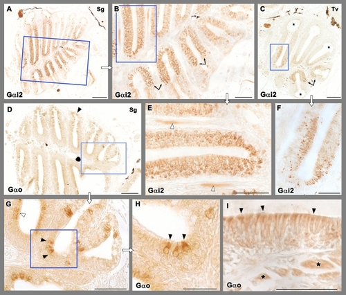
Immunohistochemical study of the olfactory rosette of zebrafish with antibodies against G-proteins. (AC,E,F) Anti-Gαi2 immunolabelling. (A) Sagittal sections show a higher number of immunopositive neurons in the internal parts of the rosette, at both sides of the median raphe. (B) Higher magnification of the inset in (A). The transition between both the nonsensory and sensory epithelium is straight (arrows). (C) Transverse sections show how the lateral rim of the lamella (asterisks) lacks of anti-Gαi2 immunolabelling. (E) Inset in (B) showing the immunopositive olfactory nerves (white arrowheads). (F) Inset in C shows how the basal part of the neuroepithelium lacks immunopositive cells. (D,GI) Anti-Gαo immunolabelling. (D) The immunoreactivity was present diffusely in apical neurons, as it is shown by open arrowheads in the inset (G). Additionally, more isolated big and oval receptor neurons showed immunoreactivity (black arrowheads) (H, inset in G). (I) The processes in the nonsensory epithelium (arrowheads) and the branches of the olfactory nerves (asterisks) are also immunopositive. Scale bar: 100 μm (A,C,D); 50 μm (B,EI).

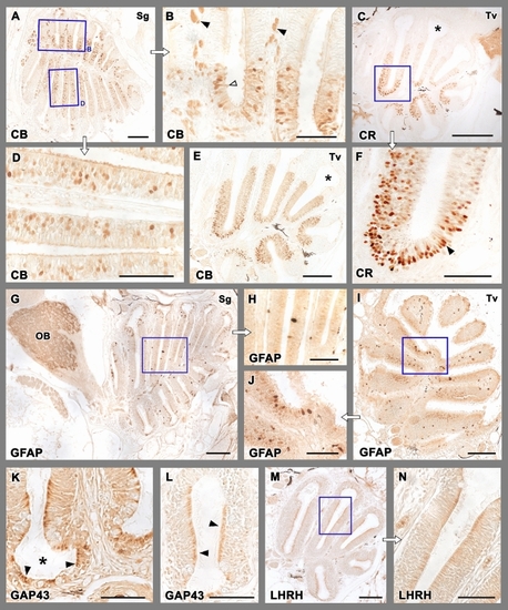
Immunohistochemical study of the olfactory rosette of zebrafish. (A,B,D,E) Anti-Calbindin labelling. (A) A sagittal section of the olfactory rosette shows labelling in a neuronal subpopulation distributed widely in the rosettes. Insets in (B) and (D) show the immunolabelling mostly located in the deeper part of the epithelium. In (B), neuroephitelial cells in the superficial layer (open arrowhead) and branches of olfactory nerves (black arrowheads) are intensely labelled. A transverse section (E) shows a similar pattern. The nonsensory epithelium (asterisk) is immunonegative. (C,F) Anti-Calretinin immunolabelling produces a stronger labelling of neuroepithelial cells, mostly concentrated in the medial part of the lamellae, and their deeper layers, whereas the nonsensory cripts (asterisk) are not immunolabelled. The neuroepithelial cells are mainly distributed in deeper layers (black arrowheads in F), and very rarely the superficial cells are lightly immunolabelled. (GJ) Anti-GFAP immunolabels isolated big cell bodies in the apical part of the epithelium. They appear in both sagittal (G) and transverse (I) sections. Insets are shown in (H) and (J), respectively. Additionally, in the olfactory bulb the antibody anti-GFAP labels the whole glomerular layer (OB). (K,L) Anti-GAP-43 immunolabelling is located in the apical part of the nonsensory epithelium (black arrowheads in K) and in individual cell bodies (black arrowheads in L) in the crypts (asterisk). (M,N) Anti-LHRH produces a light immunolabelling, mainly located in the cell processes of the nonsensory epithelium. Scale bars: 100 μm (A,CE,G,IM); 50 μm (B,F,H,N).

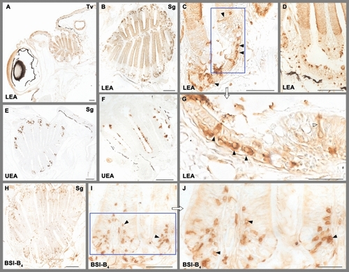
Lectin histochemical staining of the olfactory rosette. (AD,G) LEA marks intensely the olfactory epithelium dividing it clearly from the unstained nonsensory epithelium in both transverse (A) and sagittal (B) sections. In the nonsensory epithelium (C,D,G) it labels big oval neuron-like somas (black arrowheads) in the crypts. Thin dendritic processes can be clearly appreciated (white arrowhead in G). (E,F) UEA labels secretory material, mainly in the crypts, but also in the sensory epithelium. (HJ) BSI-B4 labels neuron-like cells in the nonsensory epithelium (arrowheads), including its crypts. Scale bars: 100 µm (A,B,D,E,H); 50 µm (C,F,I); 25 µm (G,J).

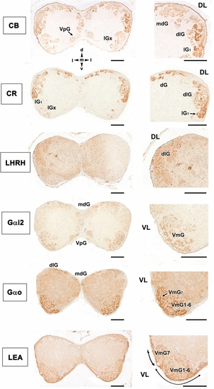
Immunohistochemical and histochemical labelling of the zebrafish olfactory bulb. Transverse sections through the central area of the olfactory bulb showing immunoreactivity with the antibodies anti-CB, anti-CR, anti-LHRH, anti-Gαi2, anti-Gαo and LEA lectin labelling. Calcium-binding proteins (CB and CR) are both mainly expressed in the dorsal (dG), dorsolateral (dlG) and lateral (lGx, lG1) glomeruli. Additionally, calbindin is also expressed in glomeruli belonging to the mediodorsal part of the bulb (mdG) and, with less intensity, in the ventral posterior area (VpG). LHRH is expressed in the dorsolateral glomeruli. Both anti G-proteins show a similar pattern, with an intense labelling in the ventromedial glomerular (VmG) area, stronger in the case of Gαo. Additionally, anti-Gαi2 marks glomeruli belonging to the ventral posterior cluster (VpG). LEA labelling is mainly circumscribed to the ventral region. DL, dorsolateral; VL, ventrolateral; dG, dorsal glomeruli; dlG, dorsolateral glomeruli; lG1, lateral glomerulus 1; lGx, lateral cluster of glomeruli; mdG, mediodorsal glomeruli; VmG1-6, ventromedial glomeruli 1–6; VmG7, ventromedial glomerulus 7; VpG, ventral posterior glomeruli; d, dorsal; l, lateral; m, medial; v, ventral. Scale bars: 100 µm.

Acknowledgments
This image is the copyrighted work of the attributed author or publisher, and ZFIN has permission only to display this image to its users. Additional permissions should be obtained from the applicable author or publisher of the image. Full text @ Sci. Rep.