
- Title
-
Therapeutic efficacy of 3,4-Diaminopyridine phosphate on neuromuscular junction in Pompe disease
- Authors
- Cinzia, B., Flavia, B., Gary, I., Renato, M., Lorenzo, M.
- Source
- Full text @ Biomed. Pharmacother.
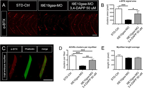
3,4-DAPP administration increased density and appropriate spatial positioning of AChRs. (A) Representative image of AChRs (α-BTX) in 4 somites of zebrafish embryos at 48 hpf collected during 3 distinct experiments. STD-Ctrl embryos: n = 32, untreated I9E10gaa-MO: n = 30, and I9E10gaa-MO treated with 3,4-DAPP 50 µM: n = 31, Scale bar = 25 µm. (B) Quantification of α-BTX-positive spots in the three experimental groups. (C) Representative image of single myofibers obtained after dissociation from 3 dpf embryos. Scale bar = 30 µm. (D,E) Quantification of AChR clusters in dissociated myofibers (D), and of myofiber average length (E) in the three experimental groups. Note that myofiber average lengths are comparable. Error bars in (B), (D), (E) are SEMs. PHENOTYPE:
|

3,4-Diaminopyridine phosphate administration increased pre- and postsynaptic signal co-localization. (A) Representative confocal fluorescence maximum projection images of SV2A (green) and α-BTX (red) signal in 5 spinal hemisegments and somites in embryos at 48 hpf. The images are representative of those found in n = 10 embryos for each experimental group (controls, morphants and 3,4-DAP treated morphants), during 3 distinct experiments. Scale bar = 25 µm. (B–D) Quantification of the SV2A (B) green signal, the α-BTX (C) red signal, and the co-localization signal (D). Error bars are SEMs. (For interpretation of the references to colour in this figure legend, the reader is referred to the web version of this article.) EXPRESSION / LABELING:
|
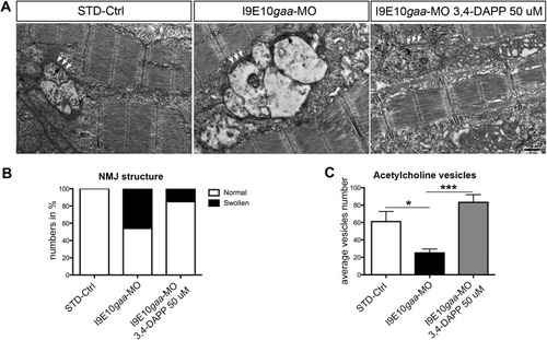
3,4-Diaminopyridine phosphate improved NMJs in zebrafish embryos. (A) Electron micrographs showing a swollen and convoluted end-plate in a morphant, a normal appearing end-plate in a 3,4-DAPP treated morphant and a normal endplate in a control embryo. Note the synaptic cleft (white arrows) and active zones (black arrows). Scale bar = 2 µm. (B,C) Quantification of the percentage of NMJs presenting normal structure (B), and of acetylcholine vesicles (C), in the three experimental groups. Error bars are SEMs. PHENOTYPE:
|

I9E10gaa-MO injected embryos behavior is ameliorated by 3,4-DAPP administration. (A, B) Representative images of 3,4-DAPP treated morphants (A), and untreated morphants (B) showing the maximum angle of flexion reached by the tail, during spontaneous tail coiling behavior at 24 hpf. (C–E) Quantification of the maximum angle of tail flexion (C), percentage of morphants showing spontaneous movements (D), and number of ipsilateral and contralateral bends of the entire body, recorded during a 30 s’ time-frame (E), in the three experimental groups. Error bars are SEMs. PHENOTYPE:
|