
- Title
-
Aquaporins Have Regional Functions in Development of Refractive Index in the Zebrafish Eye Lens
- Authors
- Wang, K., Vorontsova, I., Hoshino, M., Uesugi, K., Yagi, N., Hall, J.E., Schilling, T.F., Pierscionek, B.K.
- Source
- Full text @ Invest. Ophthalmol. Vis. Sci.
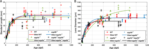
Zebrafish aqp0 mutant lens growth. Zebrafish standard length (A) and lens diameter (B) as a function of age (dpf) for WT, aqp0a−/−, aqp0b−/−, and double aqp0a−/−/aqp0b−/− mutants. WT and aqp0b−/− standard length growth plateaued at ∼24 mm after ∼350 dpf (A, red and blue); in aqp0a−/−, at ∼23 mm after ∼350 dpf (A, green). The double mutant standard length plateaued at ∼20 mm after the age of ∼180 dpf (A, black). Lens growth in WT and aqp0b−/− plateaued at ∼710 µm (B, red and blue), which corresponds to ∼500 dpf; there was no plateau for samples from aqp0a−/− mutants with a maximum attained value of 900 µm at 900 dpf (B, green), and lens growth plateaued at ∼640 µm in double homozygotes after ∼230 dpf (B, black). PHENOTYPE:
|
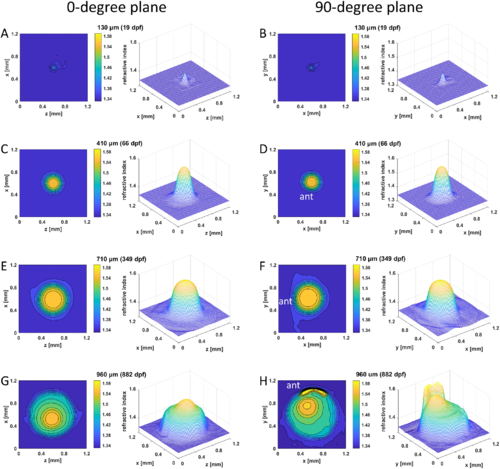
GRIN distributions in aqp0a−/− lenses. Two-dimensional contour plot (left panels) and 3D mesh plot (right panels) of GRIN distributions in aqp0a−/− lenses in the 0°plane, representing the equatorial plane (left column), and 90° plane, representing the axial plane (right column), of four selected lenses with diameters ranging from 130 to 960 µm at specified dpf. The magnitude of refractive index is color coded according to the color bar displayed on the right side of each contour plot. Anterior (ant) is the surface of the lens where light enters the lens. See Supplementary Figure S2 for higher magnifications of A and B. PHENOTYPE:
|

GRIN distributions in aqp0b−/− lenses. Two-dimensional contour plot (left panels) and 3D mesh plot (right panels) of GRIN distributions in the 0° plane, representing the equatorial plane (left column), and 90° plane, representing the axial plane (right column), of four selected lenses with diameters ranging from 130 to 790 µm at specified dpf. See Supplementary Figure S2 for higher magnifications of A and B. PHENOTYPE:
|

GRIN distributions in double aqp0a−/−/aqp0b−/− mutant lenses. Two-dimensional contour plots (left panels) and 3D mesh plots (right panels) of GRIN distributions in the 0° plane, representing the equatorial plane (left column), and 90° plane, representing the axial plane (right column), of four selected lenses with diameters ranging from 220 to 660 µm (15–713 dpf). Equatorial aspect is perpendicular to the optic axis; axial aspect contains the optic axis. Anterior (ant) is the surface of the lens where light enters the lens. See Supplementary Figure S2 for higher magnifications of A and B. PHENOTYPE:
|

Loss of Aqp0a and Aqp0b alters lens maximum refractive index growth. Lens refractive index as a function of lens diameter (A) and age (dpf) (B) for WT, aqp0a−/−, aqp0b−/−, and double aqp0a−/−/aqp0b−/− mutants. Lens maximum refractive index growth followed a similar trend and plateaued close to 1.58 at lens diameters over ∼650 µm (A) and in fish over ∼40 dpf (B). Double aqp0a−/−/aqp0b−/− mutant lens maximum refractive index growth did not plateau and reached a maximum attained value of ∼1.57 at lens diameters of ∼900 µm (A) and fish over ∼150 dpf (B). PHENOTYPE:
|
Spoke line opacity in senile WT lenses. (A, F) In vivo images of eyes of a 27-mm standard length, 1939-dpf old fish, with the corresponding dissected lens pairs illuminated by BF (B, G) and DF (C, H) illumination and the corresponding 2D contour plot (D, I) and 3D mesh plots (E, J). Spoke opacities are not visible in images taken in vivo because the iris obscures them. |

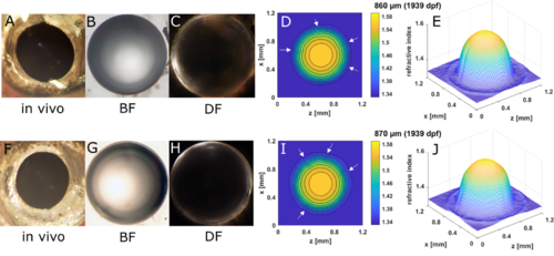
Nuclear asymmetry and anterior polar opacity in old aqp0a−/− lenses. (A, F) In vivo image of eyes of a 28-mm standard length, 676-dpf old aqp0a−/− fish, with the corresponding dissected lens pairs imaged by BF (B, G) and DF (C, H) illumination and the corresponding 2D contour plot (D, I) and 3D mesh plots (E, J) in an axial orientation. PHENOTYPE:
|
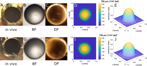
Spoke cataract in old aqp0b−/− lenses. (A, F) In vivo image of eyes of a 21.5-mm standard length, 1147-day old aqp0b−/− lens and the corresponding dissected lens pairs imaged by BF (B, G) and DF (C, H) illumination and the corresponding 2D contour plot (D, I) and 3D mesh plots (E, J). Spoke opacities are not visible in images taken in vivo because the iris obscures them. PHENOTYPE:
|

Embryonic nuclear opacity in aqp0a−/−/aqp0b−/− lenses reduced the maximum refractive index in the nucleus. (A, F) In vivo images of eyes of a 20-mm standard length, 505-dpf old aqp0a−/−/aqp0b−/− fish, with the corresponding dissected lens pairs imaged by BF (B, G) and DF (C, H) microscopy and the corresponding 2D contour plots (D, I) and 3D mesh plots (E, J). PHENOTYPE:
|

Severe opacity in aqp0a−/−/aqp0b−/− lenses reduced the overall magnitudes of GRIN profiles. (A, F) In vivo images of eyes of a 21.5-mm standard length, 713-dpf old aqp0a−/−/aqp0b−/− fish, with the corresponding dissected lens pair imaged by BF (B, G) and DF (C, H) microscopy and the corresponding 2D contour plots (D, I) and 3D mesh plots (E, J). PHENOTYPE:
|