
- Title
-
Potential of mucoadhesive nanocapsules in drug release and toxicology in zebrafish
- Authors
- Charlie-Silva, I., Feitosa, N.M., Gomes, J.M.M., Hoyos, D.C.M., Mattioli, C.C., Eto, S.F., Fernandes, D.C., Belo, M.A.A., Silva, J.O., Barros, A.L.B., Corrêa Junior, J.D., de Menezes, G.B., Fukushima, H.C.S., Castro, T.F.D., Borra, R.C., Pierezan, F., de Melo, N.F.S., Fraceto, L.F.
- Source
- Full text @ PLoS One

|

Colloidal parameters as size, PDI and zeta potential were evaluated as a function of time using the DLS technique (n = 3). All parameters were considered stable, indicating the suitability of the nanoformulations. |

The systems were maintained under magnetic stirring. Samples were collected during 2 hours and BENZ concentration was determined by HPLC. The experiments were conducted in triplicate at room temperature. The percentage of BENZ released was less than 20% for both nanocapsules, showing sustained drug release. No statistical difference was observed for the BENZ release curves (*p>0.05). |

|
|
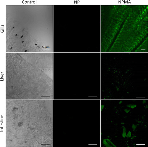
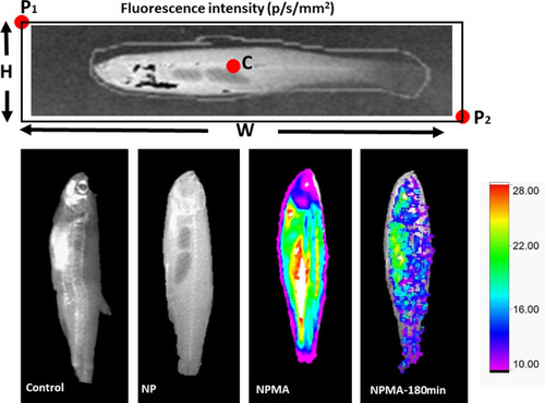
Fish gills, liver and intestine images showed fluorescence only under NPMA treatment. |
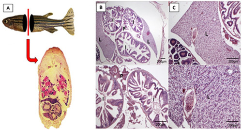
(A), (B) and (C) show fish sections without relevant pathological alterations. (L) liver; (P) pancreas and (I) intestine (H&E staining). |
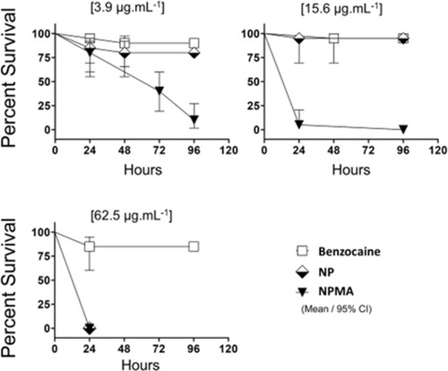
Survival rate curves of embryos exposed to BENZ, NP and NPMA at concentrations of 3.9, 15.6 and 62.5 μg/L-1. The Kaplan Meier curve was constructed and performed the log-rank (Mantel-Cox) statistical test to compare the groups. |

(A-C) control embryos presenting wildtype morphology. (D-F) Embryos exposed to BENZ revealed tail malformation (D and E) and pericardial edema (F). (G-I) Embryo contact with NP developed curved tail. (G and H) Nanoparticles formed aggregates deposition on the chorion surface. (J-L) NPMA exposure led to general malformation of embryos at 3.9 μg/L-1. |