
- Title
-
Chronic cortisol exposure in early development leads to neuroendocrine dysregulation in adulthood
- Authors
- Hartig, E.I., Zhu, S., King, B.L., Coffman, J.A.
- Source
- Full text @ BMC Res. Notes
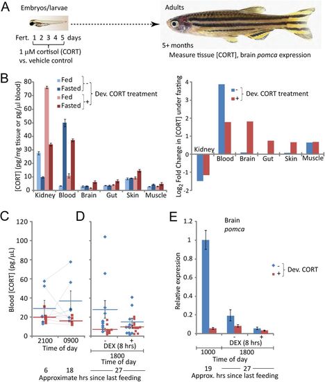
Embryos treated chronically with cortisol develop into adults with aberrant cortisol levels, tissue distribution, and dynamics. (A) Schematic of experimental design; (B) Cortisol levels in different tissues of fed and fasted (24 hours) fish (left panel) and the inferred change in those levels induced by fasting (right panel). Each measurement was taken from pooled tissues of 6 fish, with equal numbers of males and females. Error bars are the standard deviations of technical replicates. (C) Cortisol levels in blood draws from single fish at night and the following morning, with the amount of time since the last feeding indicated. Thin lines between data points indicate repeat measurements from the same fish. Averages +/− standard errors of the mean (SEM) are also shown. (D) Fasting cortisol levels in blood of individual fish (data points) following 8 hours exposure to 1 µM dexamethasone (DEX) or vehicle. The averages +/− SEM for each group are also indicated. (E) Relative expression of pomca in brain tissue in the late morning, as well as later the same day following 8 hours exposure to DEX or vehicle. The bars indicate averages +/− the standard deviation of 3 qPCR readings (technical replicates). For each measurement brain tissue was pooled from 5-6 fish, mixed males and females in equivalent proportions within each comparison group (controls 1 male and 4-5 females; cortisol-treated 2 males and 3 females). |