FIGURE SUMMARY
Title

More Favorable Palmitic Acid Over Palmitoleic Acid Modification of Wnt3 Ensures Its Localization and Activity in Plasma Membrane Domains

Authors
Azbazdar, Y., Ozalp, O., Sezgin, E., Veerapathiran, S., Duncan, A.L., Sansom, M.S.P., Eggeling, C., Wohland, T., Karaca, E., Ozhan, G.
Source
Full text @ Front Cell Dev Biol

PAM and PLM can adopt a diverse range of conformations. (A) Protein multiple sequence alignment covering a region conserved between two canonical Wnt ligands (Wnt3 and Wnt8) of vertebrates and Wingless (Wg) of Drosophila. A conserved serine amino acid (red marked column with asterisk) has been shown to be acylated in various ligands. Consensus sequences are marked in green. Dr Danio rerio, Mm, Mus musculus, Dm, Drosophila melanogaster, and Xl Xenopus laevis. (B,C) Structurally characterized (B) PAM and (C) PLM conformations downloaded from PDB (depicted in white sticks) aligned on the backbone of 4F0A's fatty acid (represented in yellow sticks). (D) The probability density distributions of being conformationally close to 4F0A's ligand. The closeness measure used is the Root-mean-square deviation (RMSD) from 4F0A's ligand, calculated over its carbon atoms. Available PLM ligand conformers have a higher probability to be <1 Å away from 4F0A's ligand. The closest ligand to 4F0A's ligand is a PLM molecule, which is depicted as an inset.

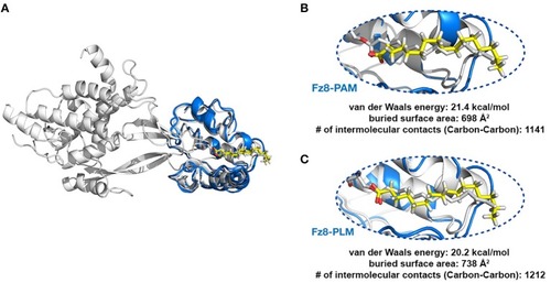
PLM modified Wnt permits conformationally viable Fz8-PLM interaction. (A) xWnt8-Fz8-PAM complex is depicted in white. xWnt8-Fz8 are represented in cartoon, where PAM is denoted in sticks. Modeled PAM and PLM ligands (including the previously missing atoms we added) are represented in yellow sticks. (B) A close-up into the Fz8-PAM interactions. Modeled PAM cannot be aligned on the oxygen atoms of 4F0A's fatty acid. (C) PLM can direct its oxygen atoms without any problems to the serine that is modified.

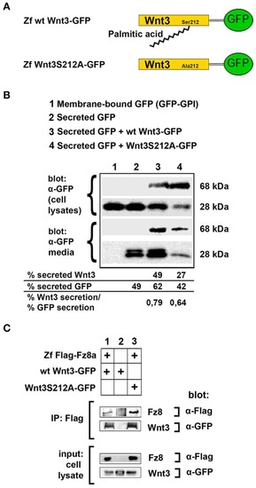
Acylation of Wnt3 at the conserved serine residue (S212) is not necessary for its secretion and interaction with its receptor. (A) Domain structure of C-terminally GFP-tagged wild-type (Zf wt Wnt3-GFP) and mutant (zf Wnt3S121A-GFP) zebrafish Wnt3. Palmitic acid is attached to the conserved serine residue. Palmitoylation is supposed not to occur in the mutant construct where serine is replaced by alanine. (B) Secretion assay for wt and mutant Wnt3 proteins and western blot of input (cell lysates) and collected media. Both wt Wnt3-GFP and Wnt3S121A-GFP are detected in the media collected from HEK293T cells transfected with corresponding constructs. Secreted and total produced levels of the proteins were calculated and used for comparisons. Membrane-bound GFP (GFP:GPI) and secreted GFP are used as negative (non-secretory protein) and positive (secretory protein) controls, respectively. Forty-nine, sixty-two, and forty-two percent of total GFP were secreted from cells transfected with secreted (sec) GFP, sec GFP+ wt Wnt3-GFP and sec GFP+ Wnt3S212A-GFP, respectively. Forty-nine and twenty-seven percent of Wnt3 were secreted from cells transfected with sec GFP+ wt Wnt3-GFP and sec GFP+ Wnt3S212A-GFP, respectively. Ratios of secreted Wnt3 to secreted GFP were 0.79 and 0.64 for wt Wnt and Wnt3S212A, respectively. Percentages represent mean of three independent experiments. (C) Coimmunoprecipitation of wt and mutant Wnt3 proteins with Frizzled8 receptor. Both wt Wnt3-GFP and Wnt3S121A-GFP coIP with Zf FLAG-Fz8a in HEK293T cells. wt Wnt3-GFP does not bind to Dynabeads non-specifically. Three independent experiments were performed.

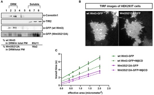
Acylation of Wnt3 facilitates its partitioning into more ordered plasma membrane environments. (A) Membrane flotation assay for wt and mutant Wnt constructs. Western blots show soluble and detergent-resistant fractions of plasma membranes derived from zebrafish embryos injected with 1 ng capped sense RNA of wt Wnt3-GFP or Wnt3S212A-GFP. DRMs and soluble fractions are marked by endogenous TfR2 and Caveolin-1 (Cav1), respectively. All Wnt proteins are detected by their GFP tags. Eighty-five percent of wt Wnt3-GFP is detected in the DRM fractions while only sixteen percent of mutant Wnt3S212A-GFP is detected in DRMs with the rest of it shifting to soluble phases. Percentages represent mean ± standard deviation (SD) of three independent experiments. (B) TIRF images of HEK293T cells expressing wt Wnt3-GFP and Wnt3S212A-GFP (C) ITIR-FCS measurements. Signal is recorded from the basolateral membrane of the cells and correlated with different bin size to form varying observation areas. The diffusion time positively correlates with the size of the area and determines the diffusion mode of the molecule as free or hindered. wt Wnt3-GFP undergoes domain-like diffusion (positive y-axis intercept) while Wnt3S212A-GFP tends to diffuse freely after cholesterol extraction. MßCD, methyl-β-cyclodextrin. Transit time and SD are obtained from three independent experiments.

Acylation of Wnt3 is essential for activation of canonical Wnt signaling. (A) Morphological phenotypes at 24 hpf of embryos injected with capped sense RNA of Wnt3-EGFP (200 pg, 53/56 embryos) or Wnt3(S212A)-EGFP (200 pg, 46/49 embryos). (B) Whole mount in situ hybridization (WMISH) showing the loss of capacity in Wnt3(S212A)-EGFP (200 pg) capped sense RNA to activate canonical Wnt signaling. First and second columns left: mCherry WMISH shows upregulation of signaling in the transgenic Tg(7xTcf-Xla.Siam:nlsm-Cherryia) canonical Wnt/ß-catenin reporter embryos by Wnt3-EGFP (36/36) but not by Wnt3(S212A)-EGFP (32/33). Second column right: Direct Wnt/ß-catenin target gene sp5l is upregulated by Wnt3-EGFP (30/32) but not by Wnt3(S212A)-EGFP (29/32). Rightmost column: Wnt3-EGFP (33/34), but not Wnt3(S212A)-EGFP (36/36), completely abolishes the telencephalon, marked by foxg1a (arrowhead), while the midbrain-hindbrain boundary, marked by her5 (arrow), is preserved. (C) Expression levels of mCherry in transgenic Tg(7xTcf-Xla.Siam:nlsm-Cherryia) and sp5l in wt embryos injected with capped sense RNAs of Wnt3-EGFP (200 pg) or Wnt3(S212A)-EGFP (200 pg) and determined by qPCR. Four independent experiments were performed. (D) Average and SD of the mean (error bars) values of pBAR luciferase reporter activity monitoring Wnt/ß-catenin signaling activity (normalized to renilla luciferase activity) in SH-SY5Y cells transfected with wt Wnt3-GFP or Wnt3S212A-GFP. wt Wnt3-GFP significantly activated the reporter whereas Wnt3S212A-GFP did not. Statistical significance was evaluated using unpaired t-test. ***p < 0.001, **p < 0.01, and n.s., non-significant. Error bars represent SD. Three independent experiments were performed.

Acknowledgments
This image is the copyrighted work of the attributed author or publisher, and ZFIN has permission only to display this image to its users. Additional permissions should be obtained from the applicable author or publisher of the image. Full text @ Front Cell Dev Biol