- Title
-
LITAF Regulates Cardiac L-type Calcium Channels by Modulating NEDD4-1 Ubiquitin Ligase
- Authors
- Moshal, K.S., Roder, K., Kabakov, A.Y., Werdich, A.A., Chiang, D.Y., Turan, N.N., Xie, A., Kim, T.Y., Cooper, L.L., Lu, Y., Zhong, M., Li, W., Terentyev, D., Choi, B.R., Karma, A., MacRae, C.A., Koren, G.
- Source
- Full text @ Circ Genom Precis Med
|
Genetic knockdown of LITAF (lipopolysaccharide-induced tumor necrosis factor) increases calcium transients in the heart of zebrafish embryos. Zebrafish hearts from 48 hpf (hours post-fertilization) wild-type (WT) and LITAF morphants (MO) were stained with Fura-2 AM (Fura-2-acetoxymethyl ester) to measure Ca2+ transients. A, Structure of human and zebrafish LITAF with conserved PXY and P(S/T)AP motifs and the hydrophobic region (HR) required for membrane embedding.49 Also indicated are 2 conserved cysteine pairs for coordination of a zinc atom likely required for proper protein folding.49 B, Averaged ventricular Ca2+ transients (amplitudes and baselines) from regions of interest (ROIs) as indicated in the color maps. C, Color maps of Ca2+ transient amplitudes from WT and LITAF morphant hearts. Color code depicts Ca2+ transient amplitudes in fluorescence ratio units (F340/F380). Squares indicate ROIs for measurements averaged in B. D, Mean baseline ratios. E, Mean Ca2+ transient amplitudes. Error bars depict SEM. One-way ANOVA, *P<0.05 for comparisons with WT (n=9 wild-type zebrafish embryos; n=7 LITAF morphants). |
|
Attenuation of Ca2+ transients and Cavα1c (L-type calcium channel alpha-1C subunit) abundance by LITAF (lipopolysaccharide-induced tumor necrosis factor) in adult rabbit cardiomyocytes. Cardiomyocytes were transduced with adenovirus expressing GFP (green fluorescence protein) or HA (hemagglutinin)-LITAF. A, Representative confocal line-scan images and corresponding Fluo-3 F/F0 time-dependent profiles at 1 Hz. B, Histograms depict mean data from Ca2+ transient amplitudes (GFP, 1.78±0.16 vs LITAF, 1.2±0.13 ΔF/F0), caffeine transient amplitudes (GFP, 2.8±0.15 vs LITAF, 2.1±0.19 ΔF/F0), fractional release and rates of Ca2+ removal by NCX (Na+/Ca2+ exchanger; kcaff), and SERCA2 (sarco/endoplasmic reticulum Ca2+-ATPase 2; kSR). Student t test, P<0.05 (2-3 heart preparations). C, Adult rabbit cardiomyocytes lysates were probed with anti-Cavα1c, anti-HA, and anti-GAPDH to indicate Cavα1c, exogenous LITAF and GAPDH (loading control) protein levels. D, Respective change in Cavα1c abundance, normalized to GAPDH (n=5 animals, performed in triplicate; mean±SEM). Student t test, P<0.05. |
|
Control of LTCC current (ICa,L) and Cavα1c (L-type calcium channel alpha-1C subunit) protein levels by LITAF (lipopolysaccharide-induced tumor necrosis factor) in 3-wk-old cardiomyocytes. Cardiomyocytes were transduced with adenovirus encoding GFP (green fluorescence protein) or HA (hemagglutinin)-LITAF for 48 h. A, Left, voltage steps to measure I-V for ICa,L. Right, representative recordings of ICa,L in cardiomyocytes. B, Voltage dependence of ICa,L current density in cardiomyocytes transduced with GFP or LITAF (cells from 5 animals; mean±SEM; Student t test, P<0.05). C, Protein levels of Cavα1c, HA-LITAF, and tubulin ( left). Respective change in Cavα1c abundance, normalized to tubulin (n=5 animals, performed in triplicate; mean±SEM). Student t test, P<0.05 ( right). D, Current-voltage relationships of ICa,L peak currents for baseline conditions from cells expressing scrambled RNA or short hairpin RNA (shRNA) against endogenous LITAF (cells from 5 animals; mean±SEM; Student t test, P<0.05). E, Protein levels of Cavα1c, total LITAF, and tubulin ( left). Respective changes in Cavα1c and LITAF abundance, normalized to tubulin (n=5 animals, performed in triplicate; right). |
|
Functional interaction between LITAF (lipopolysaccharide-induced tumor necrosis factor) and L-type calcium channel (LTCC) in tsA201 cells. Cells were transfected with plasmids for Cavα1c, (L-type calcium channel alpha-1C subunit) Cavβ3, and Cavα2δ-1 to reconstitute functional LTCC, GFP (green fluorescence protein), or HA (hemagglutinin)-tagged LITAF. Cell-surface protein levels were determined by biotinylation: cell-surface proteins were biotinylated using sulfo-NHS-SS-biotin, purified with neutravidin beads from total cell lysates, subjected to SDS-PAGE and blotted onto a nitrocellulose membrane. A, A representative Western blot shows total protein levels of Cavα1c, Cavβ3, Cavα2δ-1, HA-LITAF, and tubulin ( left). Respective change in total Cavα1c abundance, normalized to tubulin levels (n=5, performed in triplicate; mean±SEM). Student t test, P<0.05 ( right). B, A representative Western blot shows cell-surface protein levels of Cavα1c, Cavβ3, Cavα2δ-1, TFR (transferrin receptor), total LITAF, and HA-LITAF ( left). Respective changes in cell membrane protein levels of Cavα1c, Cavβ3, and Cavα2δ-1 normalized to transferrin receptor levels (n=5, performed in triplicate; mean±SEM). Student t test, P<0.05 ( right). |
|
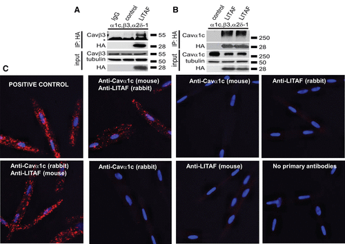
Physical interaction between LITAF (lipopolysaccharide-induced tumor necrosis factor) and L-type calcium channel (LTCC) in tsA201 cells and 3-week-old rabbit cardiomyocytes (3wRbCM). A, Immunoprecipitation (IP) of lysates from tsA201 cells transfected with plasmids for Cavα1c (L-type calcium channel alpha-1C subunit), Cavβ3, Cavα2δ-1, GFP (green fluorescence protein), or HA (hemagglutinin)-tagged LITAF using isotype control (lane 1) or HA antibody (lanes 2 and 3). A representative immunoblot against Cavβ3 shows an interaction between LITAF and the Cavβ3 subunit (IP; the asterisk indicates the heavy chain of the IP capture antibody). Also shown is the immunoprecipitated HA-LITAF protein. Input levels of Cavβ3, HA-LITAF, and tubulin are shown below. B, IP of lysates from tsA201 cells transfected with plasmids for Cavα1c, Cavβ3, Cavα2δ-1, GFP, or HA-tagged LITAF using HA antibody. A representative immunoblot against Cavα1c shows an interaction between LITAF and the Cavα1c subunit (IP). Also shown is the immunoprecipitated HA-LITAF protein. Input levels of Cavα1c, HA-LITAF, and tubulin are depicted below. C, Duo-link in situ proximity ligation assay using rabbit anti-LITAF and mouse anti-Cavα1c antibodies (alternatively mouse anti-LITAF and rabbit anti-Cavα1c antibodies) in 3wRbCM, which are amenable to proximity ligation assay and express detectable levels of LITAF and LTCC. Colocalization between molecules is indicated by red puncta. No puncta were detected in negative controls in which primary antibodies were omitted or only one antibody was used (rabbit anti-LITAF, mouse anti-Cavα1c, mouse anti-LITAF, or rabbit anti-Cavα1c antibodies). As positive control for the assay, a combination of rabbit polyclonal anti-Cavα2δ-1 and mouse monoclonal anti-Cavα2δ-1 was used to detect endogenous Cavα2δ-1. Nuclei were stained with DAPI (4',6-diamidino-2-phenylindole) (blue). |
|
LITAF (lipopolysaccharide-induced tumor necrosis factor)-mediated ubiquitination and degradation of Cavα1c (L-type calcium channel alpha-1C subunit) in tsA201 cells. Cells were transfected with plasmids for Cavα1c, Cavβ3, and Cavα2δ-1, HA (hemagglutinin)-tagged ubiquitin (HA-ubi), GFP (green fluorescence protein) as control, or Flag-tagged LITAF. Immunoprecipitation (IP) of lysates from transfected cells was performed with anti-HA antibody. A, A representative immunoblot shows levels of ubiquitinated Cavα1c ( left) and input levels of Cavα1c, Cavβ3, Cavα2δ-1, Flag-tagged LITAF, and GAPDH ( right). B, LITAF-mediated degradation of Cavα1c through lysosomes. Cells were transfected with plasmids for Cavα1c, Cavβ3, and Cavα2δ-1, GFP as control, or HA-tagged LITAF for 24 h and then treated with 10 µM chloroquine or 5 µM MG132 (N-benzyloxycarbonyl-L-leucyl-L-leucyl-L-leucinal) for 20 h. Representative Western blots show total abundance of Cavα1c and tubulin of treated cells. C, IP of lysates from cells transfected with plasmids for Cavα1c, Cavβ3, and Cavα2δ-1, HA-tagged ubiquitin, GFP as control, NEDD (neural precursor cell expressed developmentally down-regulated protein) 4-1 (N4), NEDD4-1-C867A (N4mut), or Flag-tagged LITAF was performed with anti-HA antiserum. A representative immunoblot shows levels of ubiquitinated Cavα1c ( top) and input levels of Cavα1c, Cavβ3, Cavα2δ-1, Flag-tagged LITAF, and GAPDH ( bottom). D, Respective changes in the level of ubiquitinated Cavα1c, normalized to total Cavα1c (5 experiments, performed in duplicate; mean±SEM). Student t test, P<0.05. |
|
NEDD (neural precursor cell expressed developmentally downregulated protein) 4-1-dependent downregulation of L-type calcium channel (LTCC) by LITAF (lipopolysaccharide-induced tumor necrosis factor) in 3-wk-old rabbit cardiomyocytes. A, Protein levels of total Cavα1c (L-type calcium channel alpha-1C subunit), NEDD4-1, and tubulin in cells expressing scrambled control RNA or shRNA against endogenous NEDD4-1 ( left; the asterisk indicates an unspecific band). Respective changes in NEDD4-1 and Cavα1c abundance, normalized to tubulin (n=5 animals, performed in triplicate; mean±SEM). Student t test, P<0.05 ( right). B, Current-voltage relationships of LTCC current (ICa,L) peak currents for baseline conditions from cells expressing GFP (green fluorescence protein) and short hairpin RNA (shRNA) against endogenous NEDD4-1 (control) or LITAF and NEDD4-1 shRNA. C, Three-week-old rabbit cardiomyocytes were transduced with adenovirus expressing scrambled RNA and LITAF (control) or LITAF and shRNA against endogenous NEDD4-1. Current-voltage relationships of ICa,L peak currents for baseline conditions from respective cells are depicted (cells from 5 animals; mean±SEM; Student t test, P<0.05). |
|
Computer simulation of rabbit cardiomyocytes transduced with adenovirus encoding GFP (green fluorescence protein) and LITAF (lipopolysaccharide-induced tumor necrosis factor). A, L-type calcium channel current (ICa,L) current vs clamped voltage. B– F, Current-clamp stimulation at 2.5 Hz: confocal line-scan equivalent ( B), cytosolic calcium concentration ( C), action potential ( D), ICa,L current ( E), and NCX (Na+/Ca2+ exchanger) current ( F). |


