
- Title
-
Orthogonal Drug Pooling Enhances Phenotype-Based Discovery of Ocular Antiangiogenic Drugs in Zebrafish Larvae
- Authors
- Ohnesorge, N., Sasore, T., Hillary, D., Alvarez, Y., Carey, M., Kennedy, B.N.
- Source
- Full text @ Front Pharmacol

Method overview for orthogonal drug pooling. (A) Overview of drug treatment protocol. Tg(fli1a:eGFP) positive embryos were treated from 2–5 dpf and screened for intraocular vascular defects to assess the antiangiogenic potential of test chemicals. Sunitinib was used as positive antiangiogenic control and 1% DMSO as negative vehicle control. (B) In each 96 well plate, the 8 compounds of 10 columns and the 10 compounds of 8 rows were assembled in pools (left panel). This orthogonal pooling protocol reduced 80 individual compounds to 18 test pools. Every compound is represented in two pools (right panel). (C) The primary hyaloid vessel assay readout assesses the lenses dissected (lower left and middle box) from fixed larvae and quantification of the number of primary hyaloid vessels emerging from the optic disk (asterisk) on the back of the lens counted manually under a stereomicroscope (lower right box). (D) In total, 1,760 compounds were analyzed, combined in 396 pools, resulting in one confirmed hit using this method. This assay replaces animal use with immature larval forms and the orthogonal pooling reduces the number of immature larvae needed by 45%. |

Proof-of-Principle. (A) Effect of DMSO concentration on HV assay. 1% DMSO vehicle has no effect on HV numbers. (B) The compounds of row D (D2-D11) and column 4 (4A-4H) in plate #30331 were tested individually and as pools. When 10 μM sunitinib was added to the pool “D” or “4” (pool D + Sun or pool4 + Sun), its antiangiogenic effects were as pronounced as if tested on its own (Sunitinib). (C) When compounds of plate #30251 were tested in pools, well H4 which was replaced with 10 μM sunitinib, was easily detected as antiangiogenic in both pools (“H” & “4”) containing H4. (D) A dose–response analysis with different concentrations of sunitinib alone or added to pool D in plate #30331 proves that weaker antiangiogenic effects can be reliably detected in pools. Shown are representative bright-field images of larvae, corresponding fluorescent images of dissected lenses and the quantification of primary hyaloid vessel numbers (right). Data shown are means ± SD. Statistical significance was calculated by t test and Mann Whitney test (*p < 0.05, **p < 0.01, *** p < 0.001). |

Self-deconvoluting library screen. (A) The screen of plate #30328 is shown as a representative example where 80 drugs (A2-H11) were analyzed in 18 pools (A-H, 2–11). 4 pools were lethal to the embryos (A, H, 4, 7). Three pools showed a significant reduction in blood vessel numbers, but only two (G, 9) exceeded the threshold of 40% reduction in HV numbers. (B)Individually re-tested compounds from toxic pools or of pool hits from plate #30328 identified G9 as promising candidate for further testing with >40% PHV reduction. (C) Representative images from DMSO control or 10 μM G9 treated zebrafish lenses showing the GFP-positive hyaloid vessels. (D) Three independent dose curves (test1–3) of G9 drug treatments showed strong toxicity at concentrations of >5 μM and lack of reproducible antiangiogenic activity. (E) The chemical structure of and chemical name (4-(2,4-Dichlorophenoxy)-N,N-diethyl-1-butanamine) of G9. (F) Individual re-testing of all 80 compounds from plate #30325 that were toxic in pools. No single compound reduced HV >40% and 2 were lethal at 10 μM (10G, 11B). (G) Summary of library screen. Pooled drug screens were conducted for 22 plates containing 1,760 different compounds. In 63% of cases, combinations of 8 or 10 pooled drugs did not reduce HV numbers >40%. 22% of pools caused significant developmental defects or were lethal. Of the 147 compounds identified in 18 pool hits, 80% had no significant effect when tested individually, two were toxic and eight were considered hits that significantly reduce hyaloid vessel number. Data in (A), (B), (D), and (F) are means ± SD. Statistical significance calculated by t test and Mann Whitney test (*p < 0.05, **p < 0.01, ***p < 0.001). |
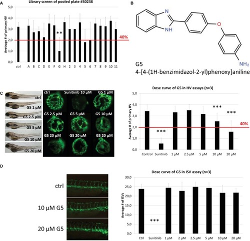
Identification and confirmation of Hit G5. (A)G5 was a promising hit identified in the screen of DIVERSet™ library plate #30238. (B) The chemical structure and chemical name (4-[4-(1H-benzimidazol-2-yl)phenoxy]aniline) of hit G5. (C) G5 shows a dose-dependent and robust reduction of primary hyaloid vessels at 10 and 20 μM. In addition, at higher doses treated larvae show no difference in survival rates and had only mild morphological changes compared to vehicle control treatments. (D) In contrast, G5 had no significant antiangiogenic effect in the ISV assay at the same range of concentrations. Data are means ± SD. Statistical significance calculated by t test and Mann Whitney test (*p < 0.05, **p < 0.01, ***p < 0.001).
|