
- Title
-
Prior Exposure to Immunosuppressors Sensitizes Retinal Microglia and Accelerates Optic Nerve Regeneration in Zebrafish
- Authors
- Bollaerts, I., Van Houcke, J., Beckers, A., Lemmens, K., Vanhunsel, S., De Groef, L., Moons, L.
- Source
- Full text @ Mediators Inflamm.
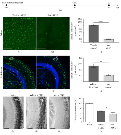
Systemic treatment with dex efficiently depletes microglia/macrophages and restricts optic nerve regeneration. (a) Schematic representation of the experimental setup. The systemic treatment with dex is started two weeks before ONC and is continued until the end of the experiment. The inflammatory response and tectal reinnervation are assessed at 6 dpi. (b-d) Prolonged systemic treatment with dex drastically reduces the number of microglia/macrophages in the retina at 6 dpi (t-test, p<0.0001). (e-g) At 6 dpi, microglia/macrophages gather in the superficial tectal layers (i.e., the stratum opticum (SO) and the stratum fibrosum et griseum superficiale (SFGS)) in vehicle-treated crushed fish. After systemic dex treatment, a similar organization of microglia/macrophages can be observed, although their number is highly reduced (t-test, p<0.05). (h-j) Representative images of biocytin-traced axons in the optic tectum of naive, uninjured fish (h) and crushed fish treated with vehicle (i) or dex (j), at 6 dpi. (k) Quantification of the regenerating RGC axons revealed that systemic immunosuppression significantly reduces tectal reinnervation at 6 dpi (t-test, p<0.05). |