
- Title
-
Fasting Upregulates npy, agrp, and ghsr Without Increasing Ghrelin Levels in Zebrafish (Danio rerio) Larvae
- Authors
- Opazo, R., Plaza-Parrochia, F., Cardoso Dos Santos, G.R., Carneiro, G.R.A., Sardela, V.F., Romero, J., Valladares, L.
- Source
- Full text @ Front. Physiol.
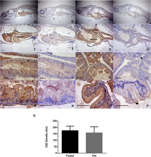
Immunohistochemical detection of ghrelin in larval zebrafish tissues in fasted and fed groups. The staining time in DAB was 30 s. Representative microphotographs were obtained at 40x magnification (A–D), 100X (E–H), 400X (I–L), and 1000X (M–P); the bars represent 50 μm. Fasted larvae are represented by photographs (A,E,I,M), contrasted with their negative controls (B,F,J,N). The fed larvae are represented by photographs (C,G,K,O), contrasted with their negative controls (D,H,L,P). Negative controls were analyzed in adjacent sections incubated without the primary antibody. The black arrows show the most intensely stained areas for the ghrelin prepropeptide, and the integrated optical density (IOD) was evaluated by the method of Paizs et al. (2009), in areas of hepatic and intestinal epithelia at 1000X microscopic magnification, using equivalent areas and ten images replicates per slide sample; the analyses was performed with Image-Pro Plus software. Bar plot (Q) show a semiquantification analysis for both study groups. Values are means ± SD (n = 6), the difference was not significant in the Mann-Whitney U-test. |