
- Title
-
Integrated one- and two-photon scanned oblique plane illumination (SOPi) microscopy for rapid volumetric imaging
- Authors
- Kumar, M., Kishore, S., Nasenbeny, J., McLean, D.L., Kozorovitskiy, Y.
- Source
- Full text @ Opt. Express
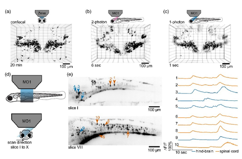
Imaging zebrafish larvae. (a) A high resolution confocal imaging of zebrafish cerebellum in nacre Tg(Olig2:GFP) fish acquired in 20 min. (b) The same cerebellar region imaged with 2P SOPi setup in 6 seconds. (c) The same cerebellar region imaged using 1P SOPi setup in 1 second. (d) Schematic diagram showing the arrangement for rapid volumetric GCaMP imaging of Tg(VGlut2a:Gal4;UAS:GCaMP6s) zebrafish hind-brain and spinal cord using a fast scanning 1P light-sheet in the SOPi setup. The volume scan consists of 10 segments, covered at rate of 100 fps leading to 10 VPS scan speed. (e) Left, GCaMP fluorescence in a subset of active cells during spontaneous activity, shown as standard deviation based intensity projections of the frames corresponding to slice position I and VIII in scanned volume (I-X). Right, GCaMP imaging traces corresponding to neurons 1-10 in optical sections I and VIII (30 sec). |