
- Title
-
Transgenic Expression of A Venous Malformation Related Mutation, TIE2-R849W, Significantly Induces Multiple Malformations of Zebrafish.
- Authors
- Du, Z., Ma, H.L., Zhang, Z.Y., Zheng, J.W., Wang, Y.A.
- Source
- Full text @ Int J Med Sci
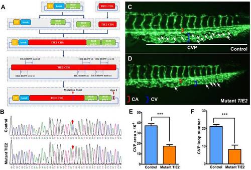
Construction of plasmid encoding humanized TIE2-R849W and CVP defect in zebrafish caused by TIE2-R849W expression. (A) Construction of plasmid encoding mutant TIE2, based on normal humanized TIE2 sequence, via PCR-mediated site-directed mutagenesis. Location of mutation-related primers and plasmid elements are indicated. (B) Traditional gene sequencing identification after construction. As shown by red arrow, nucleotide T in control group had successfully transformed to C in mutant TIE2 group. (C) At approximately 2 days after fertilization (dpf) in control embryos, CVP formed honeycomb-like structures at the tail (white arrowheads). In contrast, mutant human TIE2 mRNA (200 pg) injection resulted in specific defects in caudal vein plexus (CVP) formation (D, white arrowheads). (E, F) Quantification of area and loop formation at CVP (Scale bars are 100 μm for A-C. Error bars, s.e.m.; *** refers to P < 0.0001 by ANOVA. CVP, caudal vein plexus; CA, caudal artery; CV, caudal vein). |

Obvious eye defects of zebrafish caused by humanized TIE2-R849W. (A) At 52 hpf, compared to normal embryogenesis of eyes in the control group, from the angle of dorsal view and ventral view, there exist apparently unilateral or bilateral eyelessness, reduction of size in one side and eye-fusion at dorsal position (impaired eyes are indicated by blue arrow). (B) Quantification of eye defects in control and TIE2-R849W group at 52 hpf. (C) Histological research (HE) on reduced size eye (7 dpf) indicates loss of lens (indicated by green asterisk), abnormal plexiform layer (indicated by yellow asterisk), and still-recognizable separated layer structure (showed by partial enlargement). (HE, hematoxylin and eosin staining; scale bars are 100 μm for C.) |

TIE2-R849W-related malformed forebrain and abnormal jaw developmental extension in zebrafish. (A, B) Injection of TIE2-R849W mRNA causes malformed forebrain and abnormal jaw developmental extension by 7 dpf. There is a slightly different but almost similar degree of alteration among different individuals. (C, D) From the profile view, despite the obvious perturbations in the forebrain (indicated by white asterisk) and mandibular structures (indicated by blue triangle), the otocysts develop normally (indicated by orange arrow). (E, F) In the mutant group, there exists mandibular hypermorphosis towards the anterior inferior part, reduction of mandibular width, holistic morphological abnormalities and mild bimaxillary deviation. (G) Quantification of jaw developmental defects in control and TIE2-R849W group at 52 hpf. (H, I) In contrast to the control group (I), zebrafish in the TIE2-R849W group were histologically characterized by abnormal extension of mandibular cartilage (indicated by green arrow), ectopic proliferation of myelencephalon (indicated by orange arrow) and aberrant location of neurogliocytes (indicated by black asterisk) (HE, hematoxylin and eosin staining; scale bars are 100 μm for H and I). |