
- Title
-
Spinal cholinergic interneurons differentially control motoneuron excitability and alter the locomotor network operational range
- Authors
- Bertuzzi, M., Ampatzis, K.
- Source
- Full text @ Sci. Rep.

Organization of spinal cholinergic system. (A) Distribution of cholinergic terminals within the adult zebrafish spinal cord. (B) Single optical sections show the cholinergic synapses onto different types of motoneurons (MNs). (C) Cholinergic (ACh) input is different between motoneuron (MN) classes, (nslow = 10, ninterm = 27, nfast = 30 neurons; n = 5 zebrafish). Quantification of inputs on each MN obtained from a single focal plane images. (D) Whole mount spinal cord immunohistochemistry reveals that a fraction of ChAT+ neurons are not backfilled motoneurons. Arrows indicate ChAT+MN− neurons (Cholinergic interneurons). (E) Quantification of the adult zebrafish cholinergic neurons, per spinal cord hemisegment, that are motoneurons or interneurons (n = 7 zebrafish). (F) Cholinergic interneurons (ChATINs) have smaller soma sizes compared to motoneurons (ChATMNs). (G) Representative example of the distribution pattern of cholinergic interneurons (ChATINs) and motoneurons (ChATMNs) in the adult zebrafish spinal cord. (H) Injection of a retrograde dextran tracer in spinal segment 18 reveals the descending interneurons located in spinal cord segment 15. Few descending neurons that are cholinergic (ChAT+) were found in the spinal hemisegment of adult zebrafish. Arrow indicates cholinergic descending interneuron. (I) Injection of dextran retrograde tracer in spinal segment 12 reveals the ascending interneurons in spinal cord segment 15. Arrows indicate a small number of retrograde traced neurons that are cholinergic interneurons. (J) Analysis of the number of the cholinergic interneurons that possess a descending (2.28 ± 0.28 neurons/hemisegment, n = 7 zebrafish) or an ascending (4 ± 0.3 neurons/hemisegment, n = 7 zebrafish) axon. (K) The descending and ascending cholinergic interneurons have non-overlapping soma sizes (t = 13.4, p < 0.0001, n = 37 neurons), suggesting that different populations of cholinergic interneurons are ascending or descending in adult zebrafish spinal cord. (L) Percentage of the descending and ascending cholinergic interneurons per hemisegment of spinal cord. CC, central canal; D, dorsal; L, lateral; MA, Mauthner axon; P, posterior; V, ventral. Data are presented as mean ± SD; ***p < 0.001; ****p < 0.0001; n.s., non-significant. |
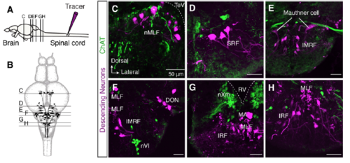
Brain neurons descending to spinal cord are not cholinergic. (A-B) Injection of methylrhodamine dextran in the spinal cord retrogradely labels all the descending supra-spinal neurons (N = 8 zebrafish brains). Schematic representation of the distribution of adult zebrafish brain descending neurons with the level of sections that correspond to the following images in C-H. (C-H) Confocal images show that none of the descending labeled neuron is ChAT+ in all studied brain areas. DON, descending octaval nucleus; IMRF, intermediate reticular formation; IRF, inferior reticular formation; MA, Mauthner axon; MLF, medial longitudinal fascicle; nMLF, nucleus of the medial longitudinal fascicle; nVI, abducens nucleus; nXm, vagal motor nucleus; RV, rhombencephalic ventricle; SRF, superior reticular formation; TeV, tectal ventricle. |

m2-mAChRs in different motoneurons. (A-B) Distribution pattern of m2-mAChRs in relation to different motoneuron types. A combination of retrograde labeling of different motoneuron pools with immunostaining for m2-mAChRs reveals that the fast motoneurons exhibit a vast number of m2 receptors compared to other motoneuron types (intermediate and slow). Slow motoneurons were found to be weakly labeled. Data are presented as mean ± SD; *p < 0.05; ****p < 0.0001. |
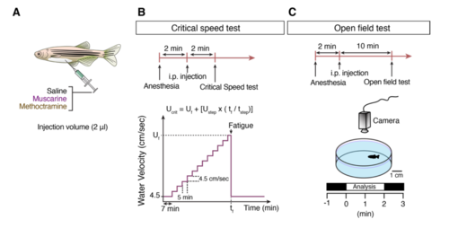
Experimental design to investigate the effect of mAChRs during in vivo locomotion. (A) Intraperitoneal administration of saline, muscarine and/or methoctramine in anesthetized adult zebrafish before the in vivo tests. (B) The critical speed test protocol. (C) Protocol for in vivo monitoring of adult zebrafish locomotor behavior. |