
- Title
-
Overexpression and Knockdown of Hypoxia-Inducible Factor 1 Disrupt the Expression of Steroidogenic Enzyme Genes and Early Embryonic Development in Zebrafish
- Authors
- Tan, T., Yu, R.M.K., Wu, R.S.S., Kong, R.Y.C.
- Source
- Full text @ Gene Regul Syst Bio
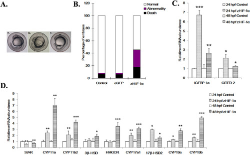
Effects of zHIF-1α overexpression on embryonic development and the expression of hypoxia markers and steroidogenic genes. (A) Development of zebrafish embryos at 24 hpf. a, uninjected control embryo; b, embryo microinjected with eGFP mRNA shows normal development (Prim-6 stage); c, embryo microinjected with zHIF-1α-ΔODD-eGFP mRNA shows developmental retardation (at approximately 18-somite stage). (B) Percentage of mortality and morphological abnormality in zebrafish embryos (24 hpf) microinjected with zHIF-1α mRNA. The effect of zHIF-1α overexpression on steroidogenic gene expression was examined by microinjecting zHIF-1α mRNA into 1- to 2-cell stage zebrafish embryos and maintained under normoxic conditions. Zebrafish embryos microinjected with eGFP mRNA and maintained under normoxia were used as the control. Because no statistically significant difference was observed between the data sets of the microinjected control and the no-injection control (One-way analysis of variance with a P < .05 threshold), the latter data set is presented here. Control: uninjected embryos (n = 373, dead = 31, abnormal = 4); eGFP: embryos microinjected with eGFP mRNA (n = 719, dead = 48, abnormal = 16); and zHIF-1α: embryos microinjected with zHIF-1α-ΔODD-eGFP mRNA (n = 1081, dead = 193, abnormal = 297). (C) Effects of zHIF-1α overexpression on IGFBP-1a and CITED-2 expression. (D) Effects of zHIF-1α overexpression on steroidogenic gene expression. Gene expression was quantified using real-time PCR and normalized with β-actin mRNA. Data are presented as the mean relative fold change ± SD with respect to the gene expression level in the control (its expression level was arbitrarily set to 1) for each experiment. Expression levels significantly different from the control are indicated by asterisks (t test, n 4, *P .05, **P .01, ***P .001). eGFP indicates enhanced green fluorescent protein; mRNA, messenger RNA. |

Effects of zHIF-1α knockdown on zebrafish embryonic development and the expression of hypoxia markers and steroidogenic genes under normoxia. (A) Morphological abnormalities of the zHIF-1α knockdown embryos at 48 hpf under normoxia. The effect of zHIF-1α knockdown on steroidogenic gene expression was examined by microinjecting zHIF-1α MOs into 1- to 2-cell stage zebrafish embryos and maintained under either normoxia or hypoxia. Zebrafish embryos microinjected with a standard control MO were used as controls. All morphants were imaged at the left lateral view. a, Control: embryo injected with control MO; b-j, embryos microinjected with zHIF-1α MO; b, c, and d, morphants with decreased body length, bended notochords (black arrows), and distorted abdomens (blue arrows); e-j, morphants with bended notochords, small head circles (red circles), and short curved tail (yellow arrows). (B) Effects of zHIF-1α knockdown on IGFBP-1a and CITED-2 expression under normoxia. (C) Effects of zHIF-1α knockdown on steroidogenic gene expression under normoxia. Gene expression was quantified by real-time PCR and normalized against β-actin mRNA. Data are presented as the mean relative fold change ± SD with respect to the gene expression level in the control (its expression level was arbitrarily set to 1) for each experiment. Expression levels significantly different from the control are indicated by asterisks (t test, n 4, *P .05, **P .01, ***P .001). mRNA indicates messenger RNA; MO, morpholino oligonucleotide. |