
- Title
-
A Simple Predictive Enhancer Syntax for Hindbrain Patterning Is Conserved in Vertebrate Genomes
- Authors
- Grice, J., Noyvert, B., Doglio, L., Elgar, G.
- Source
- Full text @ PLoS One
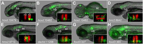
CNEs containing PBX-HOX motifs driving expression in hindbrain of transgenic zebrafish. Images show expression driven during transient transgenic assays (see methods) between 2 and 3 dpf. Insets show comparison with RFP in rhombomeres 3 and 5. hoxd.10479 (A) at ~42 hpf in ventral r5r6; hoxd.10482 lateral (B) and dorsal (C) views at ~56 hpf in lateral r2, r4, r6, pectoral fin and spinal cord; bnc2.8642 (D) at ~60 hpf in hindbrain; hmx2.9713 (E) at ~60 hpf in hindbrain and spinal cord; dachd.11206 (F) at ~72hpf in hindbrain and spinal cord; foxd3.327 (G) at ~72hpf in ventral r5 and r6; foxd3.365 throughout the central nervous system, most strongly in the ventral hindbrain, and cranial ganglia. fb: forebrain; mb: midbrain; hb: hindbrain; sc: spinal cord; pf: pectoral fin; pa: pharyngeal arches/neural crest; cg: cranial ganglia; r3 r5: rhombomeres 3 and 5. |
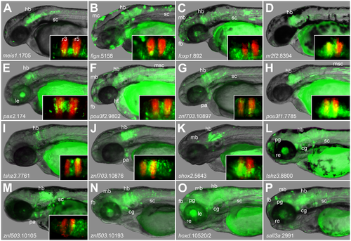
Conserved PBX-HOX and MEIS/PREP motifs predict hindbrain enhancers accurately. Images show expression driven during transient transgenic assays (see methods) between 2 and 3 dpf. Insets show comparison with RFP in rhombomeres 3 and 5. Constructs drive expression in hindbrain and in other tissues meis1.1705 (A) in hindbrain and spinal cord; fign.5158 (B) in hindbrain, spinal cord and melanocytes; foxp1.892 (C) in the central nervous system; nr2f2.8394 (D) in hindbrain; pax2.174 (E) in hindbrain and lens; pou3f2.9802 (F) in the central nervous system, heart and muscle; znf703.10897 (G) in hindbrain, spinal cord and pharyngeal arches/neural crest; pou3f1.7785 (H) in hindbrain; tshz3.1.7761 (I) in hindbrain; znf703.10876 (J) in hindbrain and pharyngeal arches/neural crest; shox2.5643 (K) in hindbrain; tshz1.8800 (L) in hindbrain, spinal cord, retina, pineal gland and cranial ganglia; znf503.10105 (M) in hindbrain, spinal cord and pharyngeal arches; znf503.10193 (N) in the central nervous system and cranial ganglia; hoxd.10520/1 (O) in the central nervous system, lens, retina, pineal gland and cranial ganglia; and sall3a.2991 (P) in the central nervous system, retina, pineal gland and cranial ganglia. fb: forebrain; mb: midbrain; hb: hindbrain; sc: spinal cord; pa: pharyngeal arches/neural crest; cg: cranial ganglia; mc: melanocytes; msc: trunk muscle cells; le: lens; re: retina; pg: pineal gland. |
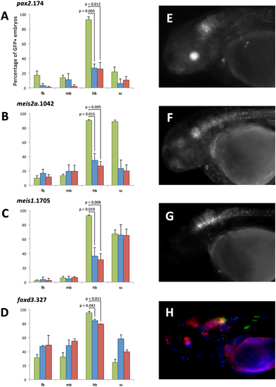
The PBX-HOX and MEIS/PREP motifs of four enhancers are essential for their function. Histograms for four elements, pax2.174 (A), meis2a.1042 (B), meis1.1705 (C) and foxd3.327 (D), showing the number of embryos with GFP positive cells in forebrain (fb), midbrain (mb), hindbrain (hb) and spinal cord (sc) when expressing wild-type (green), MEIS/PREP site mutant (blue) or PBX-HOX site mutant (red) constructs. Annotation displays p values for one-tailed paired t tests. All mutant constructs show a significant (student's t test p = <0.05) reduction in the number of embryos positive for hindbrain. Wild-type pax2.174 (E) drives expression in hindbrain and lens (green), whereas mutant constructs do not drive this pattern. Wild-type meis2a.1042 (F) drives expression in the central nervous system particularly the anterior hindbrain. Mutant constructs fail to recapitulate this expression. Wild-type meis1.1705 (G) drives expression in the hindbrain and spinal cord, but mutant constructs drive expression only in spinal cord. Wild-type foxd3.327 (H) drives expression in posterior hindbrain (green), but expression driven by constructs where either the MEIS/PREP (blue) or the PBX-HOX (red) motif is mutated is frequently ectopic in midbrain, anterior hindbrain and spinal cord. |
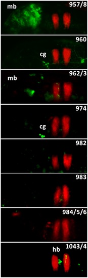
Enhancer activity driven by meis2a CNEs containing PBX-HOX or MEIS/PREP motifs. Images show the hindbrain and midbrain region of 48 hpf zebrafish embryos. GFP shows enhancer activity driven by injected constructs. RFP shows hindbrain rhombomeres 3 and 5. cg: cranial ganglia; hb: hindbrain; mb: midbrain. |