
- Title
-
Deep Brain Photoreceptor (val-opsin) Gene Knockout Using CRISPR/Cas Affects Chorion Formation and Embryonic Hatching in the Zebrafish
- Authors
- Hang, C.Y., Moriya, S., Ogawa, S., Parhar, I.S.
- Source
- Full text @ PLoS One
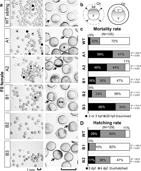
Abnormal chorion elevation in the eggs or embryos obtained from F0 female valopa/b mutants. (a) Images of zebrafish eggs or embryos obtained at 4 hour-post-fertilization (hpf) from a female WT sibling and female mutants: valoparw01 (represented by A1), valoparw02 (A2), valopbrw01 (B1), valopbrw02 (B2), valopbrw03 (B3). Upon spawning, chorion is elevated from the surface of the egg, and the blastodisc develops into embryonic cells in the created space (b). Arrows indicate the chorion surrounding the egg. Note that the eggs or embryos obtained from the valopa/b mutants exhibited no (single asterisk) or only partial (double asterisks) chorion elevation. Eggs with partial chorion elevation exhibited blastodisc expansion; although some develop into embryos, all eventually died (opaque appearance). (c and d) The mortality rates of the eggs or embryos and the hatching rates of embryos, respectively. Embryos from B1 and B2 mutants survived but mostly failed to hatch at 4 day-post-fertilization (dpf), in contrast to most embryos from the WT sibling hatched. Note that the association between the valop mutations in individual F0 parents and the increased number of dead eggs/embryos at 26 hpf and the decreased number of hatched embryos at 4 dpf was statistically significant (determined by Chi-Square X2 values and p-values). Logistic Regression analysis showed that the observed phenotype is likely dependent on the targeted mutation (data in S1 File). N represents the number of eggs or embryos; and percentages shown in a single bar add up to a total of 100%. Abbreviation: bd, blastodisc; c, embryonic cells; Ch, chorion; y, yolk. Scale bar, 1 mm. |

Late-hatching embryos obtained from a F0 male valopa mutant. (a) Images of the zebrafish embryos obtained from a male WT sibling and a male mutant valoparw03 (represented by A3) at 0 day-post-fertilization (dpf), 4 dpf, and 6 dpf. (b and c) the mortality rates of the eggs or embryos and hatching rates of the embryos, respectively. Most embryos from the mutant had not hatched at 4 dpf, in contrast to most embryos from the WT sibling hatched. At 6 dpf, most of the unhatched embryos had died in the chorion. Genotyping of F1 adult offspring identified a mixture of WT and mutants. Note that the association between the valop mutations in individual F0 parents and the decreased number of hatched embryos at 4 dpf was statistically significant (determined by Chi-Square X2 values and p-values). Logistic Regression analysis showed that the observed phenotype is likely dependent on the targeted mutation (data in S1 File). N represents the number of eggs or embryos; and percentages shown in a single bar add up to a total of 100%. Scale bar, 1 mm. PHENOTYPE:
|

Unillustrated author statements PHENOTYPE:
|