
- Title
-
Production of reproductively sterile fish by a non-transgenic gene silencing technology
- Authors
- Wong, T.T., Zohar, Y.
- Source
- Full text @ Sci. Rep.
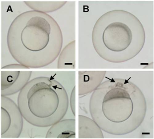
Vivo conjugated Morpholino oligomer (MO) caused developmental delays and uncharacterized aggregates in zebrafish embryos. When treated with 20 µM dnd-MO, embryos developed normally and reached (A) 1K cell stage after a 3 hour immersion and (B) 30-50% epiboly stage after a 5 hour immersion. When treated with 20 µM Vivo conjugated dnd-MO (dnd-MO-Vivo), embryo development was slightly delayed and only reached (C) 256-512 cell stage with aggregates found between the chorion and blastodisc (arrows) after a 3 hour immersion, and only reached (D) sphere stage with more aggregates (arrows) after a 5 hour immersion. Scale bar = 200 µm.
|
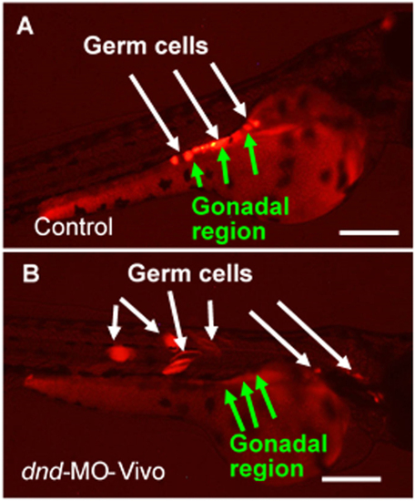
Zebrafish dnd-MO-Vivo disrupted germ cell development in zebrafish. (A) In control embryos immersed in only water or dnd-MO solution, germ cells migrated to the gonadal region and maintained their morphology as round-shaped cells. (B) Treatment of dnd-MO-Vivo caused germ cell mis-migration and eventually differentiation into other cell types that can be clearly seen by the change of their morphology. Scale bar = 200 µm. |
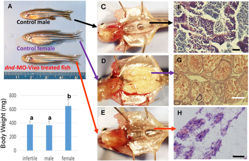
Zebrafish dnd-MO-Vivo induced sterility in zebrafish. Embryos initially treated with 60 or 40 µM of dnd-MO-Vivo developed into male-like adults. (A) No difference in appearance or overall size was observed between treated adult fish and control males. (B) No significant difference in body-weight (Mean ± SD) of 3-month-old fish (N = 12 by random sampling) was noted among dnd-MO-Vivo treated fish and control males (Data that share the same letter are not significantly different from each other). Examination of gonadal tissue showing (C) a fully-developed testis of a control male fish, (D) a fully-developed ovary of a control female fish, (E) the gonads of dnd-MO-Vivo treated fish that developed into a thin filament-like tissue. Photomicrographs (F–H) show (F) advanced spermatogenesis in the testis of a control male fish, (G) a well-developed ovary of a control female fish with oocytes at advanced stages of gametogenesis, (H) the gonad of dnd-MO-Vivo treated fish appears to be under-developed and surrounded with a large amount of adipocytes, without advanced gonadal structure or germ cells. Scale bar: white = 200 µm, black = 20 µm. PHENOTYPE:
|