
- Title
-
Are zebrafish larvae suitable for assessing the hepatotoxicity potential of drug candidates?
- Authors
- Mesens, N., Crawford, A.D., Menke, A., Hung, P.D., Van Goethem, F., Nuyts, R., Hansen, E., Wolterbeek, A., Van Gompel, J., De Witte, P., Esguerra, C.V.
- Source
- Full text @ J. Appl. Toxicol.

Different lfabp10a expression patterns observed in 6 days post fertilization larvae. (A) Normal expression. (B) Reduced expression. (C) Reduced size. (D) Absent expression. (E) Enlarged size. |

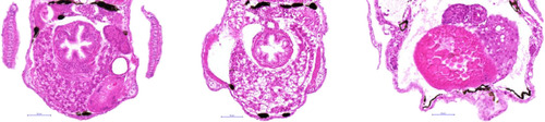
Hepatocellular glycogen variation in control livers. Histopathological analysis of transversal HE sections of control larvae showing varying levels of glycogen accumulation from no observable accumulation (A), to minimal (B), very mild (C) and mild accumulation (D). Yellow border indicates the liver. Magnification 400×. |
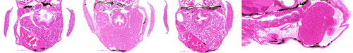
(A) Transversal H&E liver sections of a control larva and a Diclofenac-treated larva showing moderate accumulation of glycogen in the liver (B) or a small liver (C) with hypertrophic, vacuolated hepatocytes (fat arrows). Yellow border indicates the liver. (D) Detailed image from (C) showing the vacuolization. Magnification: 400×. |

Transversal H&E liver section of a control larva (A) and Troglitazone-treated larva showing a small liver (B) with hypertrophic, vacuolated hepatocytes (fat arrows), in combination with edema (thin arrows). Saggital section of a Troglitazone-treated larva showing a small liver (C) with hypertrophic, vacuolated hepatocytes (fat arrow), in combination with edema (thin arrows). Yellow border indicates the liver. Magnification: 400×. |

Transversal H&E liver slides of a control larva (A) and a Rosiglitazone-treated larva (B). Hepatocytes in the liver of the Rosiglitazone-treated larva are normal and comparable to the controls. (C) Saggital H&E section of a Rosiglitazone-treated larva. No hepatocellular abnormalities are observed. |
Transversal H&E liver section of a control larva (A) and an Alpidem-treated larva showing enlarged livers with hepatocellular hyperplasia (B) or hypertrophic vacuolated hepatocytes (C). Saggital H&E section of an Alpidem-treated larva showing an enlarged liver with hepatocellular hyperplasia and edema (arrows) (D). Yellow border indicates the liver. Magnification: 400×. |