
- Title
-
Molecularly defined unfolded protein response subclasses have distinct correlations with fatty liver disease in zebrafish
- Authors
- Vacaru, A.M., Di Narzo, A.F., Howarth, D.L., Tsedensodnom, O., Imrie, D., Cinaroglu, A., Amin, S., Hao, K., Sadler, K.C.
- Source
- Full text @ Dis. Model. Mech.

Tm is the only ER stressor that induces fatty liver. (A,B) Representative images of 5-dpf larvae treated for 48 hours with DMSO, BFA, Tg or Tm at the indicated concentrations. (A) Live transgenic Tg(fabp10:dsRed) larvae were imaged. (B) Fixed larvae were stained for neutral lipids with Oil Red O. Livers are circled. (C) Larvae treated with DMSO, 1 and 2 μg/ml BFA, 0.25, 0.5 and 0.75 μM Tg or 1 µg/ml Tm from 3-5 dpf were stained with Oil Red O and scored for the presence (black bars) or absence (white bars) of steatosis. The total number of larvae scored and the number of clutches are indicated. **P<0.01, ***P<0.0001, calculated using Fisher’s exact test. Scale bars: 200 μm (A), 1 mm (B). EXPRESSION / LABELING:
|
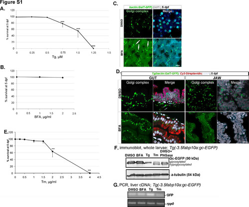
Tg, BFA and Tm have different toxicities and affect differentially the secretory pathway morphology and function. A. Tg causes dose-dependent lethality of zebrafish larvae exposed from 3 to 5 dpf. B. Survival of zebrafish larvae treated with 1 and 2 μg/ml BFA from 3-5 dpf. C. Percent of larvae that survive exposure to Tm concentrations ranging from 0 to 4 μg/ml from 3 to 5 dpf was scored in at least 3 clutches. The maximal tolerable dose was determined as 1 μg/ml. D. Lysates of single 5 dpf Tg(l-fabp:gc- EGFP) larvae treated with DMSO, 1 mg/ml BFA, 0.75 μM Tg or 1 mg/ml Tm were immunoblotted with anti-GFP and anti-a-tubulin to detect a mobility shift in Gc-EGFP representing a change in glycosylation status. +PNGase indicates that protein lysates were treated with PNGase. E. PCR analysis of GFP and rpp0 from samples treated as in D. F. Confocal images of sections of hepatocytes from Tg(bactin:GalT-GFP) larvae treated with DMSO and 1 mg/ml BFA. Nuclei are stained with DAPI (blue). Scale bar = 10 μm. F. Confocal images of the gut and jaw from cryosections of transgenic 5 dpf Tg(bactin:GalT-GFP) zebrafish larvae treated from 3 to 5 dpf with DMSO (upper panels) or with 1 mg/ml BFA (lower panels) show a complete dispersal of the GC in case of the BFA treatment as compared with the DMSO control. Sections were stained with Cy3-streptavidin (red) to visualize the gut. |