
- Title
-
Ethanol affects the development of sensory hair cells in larval zebrafish (Danio rerio)
- Authors
- Uribe, P.M., Asuncion, J.D., and Matsui, J.I.
- Source
- Full text @ PLoS One
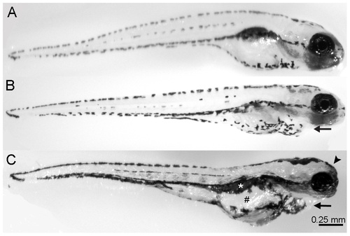
Ethanol treatment affects the development of larval zebrafish. Zebrafish embryos were raised in varying concentrations of ethanol (0% through 1.5% by volume) from 2 days post-fertilization (dpf) to 5 dpf, fixed, and imaged. (A) Untreated control animals appear normal. (B) 1% ethanol-treated animals have a slightly swollen heart (arrow) and no swim bladder but otherwise appear normal. (C) Increasing the concentration of ethanol treatment to 1.5% resulted in embryos exhibiting swollen hearts (arrow), swollen guts (#), missing a swim bladder (white asterisk), and inhibited craniofacial development (arrowhead), which is similar to the phenotype exhibited by children with FAS. PHENOTYPE:
|
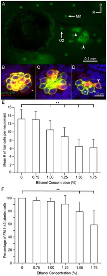
Ethanol but not methanol treatment affects the number of functional sensory hair cells in the lateral line. Brn3c-GFP transgenic zebrafish embryos were raised in embryo medium supplemented with varying concentrations of ethanol. Prior to fixation, the larvae were stained with FM 1-43FX (red) to determine if hair cell mechanotransduction channels were functional. (A) Lateral view of a Brn3c-GFP zebrafish. Clusters of bright green GFP-labeled hair cells are found in neuromasts along the head and body of the animal. The O2 and Mi1 neuromasts are highlighted along with inner ear organs (small arrows). Higher magnification of hair cells of the O2 neuromast taken from z-stacks show that (B) hair cells in untreated controls and (C) 1% ethanol-treated animals had no observable morphological differences though there were fewer hair cells in the 1% ethanol-treated animals. Almost all of the GFP-labeled hair cells were co-stained with FM 1-43FX. (D) Fewer sensory hair cells were observed in neuromasts of animals treated with 1.5% ethanol. There were also fewer double-labeled cells (arrowhead) and more single labeled cells (asterisk). (E) Stereocilia bundles were counted to determine the number of hair cells present in the O2 neuromast. Control numbers were similar to those reported in other studies [23], [31]. Significantly fewer hair cells (**p<0.01) were counted in ethanol-treated larvae at concentrations greater than 1% per volume when compared to controls. No differences in the number of hair cells were observed in larvae treated with 1.5% methanol. (F) The percentage of GFP-labeled cells that were also co-stained for FM 1-43 decreased as the concentration of ethanol increased but not in methanol-treated larvae. There was a significant decrease (**p<0.01) in the number of double-labeled hair cells at the two highest concentrations of ethanol tested. Results are the mean values ± SD. n = 8-21 per condition. |
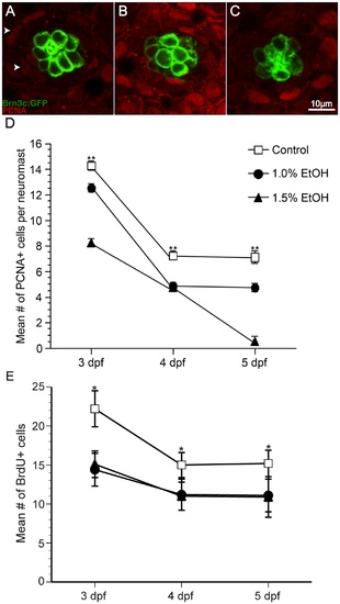
Ethanol exposure significantly reduced the number of proliferating cells compared to untreated controls in O2 neuromasts of Brn3c GFP-larvae. (A) Under untreated control conditions, proliferating cell nuclear antigen (PCNA)-labeled cells (arrowhead) are primarily non-sensory supporting cells and mantle cells in the neuromast (area demarked by white dotted line). Fewer PCNA-labeled cells result from ethanol treatment at (B) 1% and (C) 1.5% by volume. (D) The mean number of PCNA-labeled cells decreases during maturation and according dose of ethanol. (E) In a separate experiment, bromodeoxyuridine was added to the embryo medium during the last 24 hours of treatment, fixed, processed for BrdU immunohistochemistry, and BrdU-labeled cells were counted. Significantly fewer BrdU-labeled cells were observed in ethanol-treated animals when compared to untreated controls. Results are the mean values ± SD. n = 10-21 neuromasts for each treatment group. **p<0.01. EXPRESSION / LABELING:
PHENOTYPE:
|
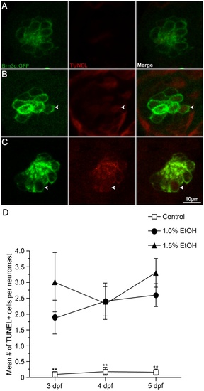
Ethanol exposure significantly increased the number of TUNEL-labeled cells in the O2 neuromast. Apoptotic activity in O2 neuromasts was measured by TUNEL labeling of untreated and ethanol-treated Brn3c-GFP larvae from 2-5 dpf. (A, D) Untreated controls showed few TUNEL-labeled cells but (B, D) 1% ethanol-treated and (C, D) 1.5% ethanol-treated larvae showed a significant increase in the number of TUNEL-labeled cells (arrowhead) versus control animals. While there were more TUNEL-labeled cells in ethanol-treated animals when compared to untreated controls, there was no statistical difference between ethanol concentrations. Results are the mean values ± SD. n = 9-12 neuromasts for each treatment group. *p<0.05 and **p<0.01 when compared to untreated controls. EXPRESSION / LABELING:
PHENOTYPE:
|