
- Title
-
Developmental expression pattern of two zebrafish rxfp3 paralogue genes
- Authors
- Fiengo, M., Del Gaudio, R., Iazzetti, G., Giaimo, R.D., Minucci, S., Aniello, F., and Donizetti, A.
- Source
- Full text @ Dev. Growth Diff.

Reverse transcription-polymerase chain reaction (RT-PCR) on rxfp3-2a and rxfp3-2b transcripts during zebrafish embryogenesis. Temporal expression pattern of zebrafish rxfp3-2a and rxfp3-2b at different embryonic stages indicated on top as hours post fertilization. Amplification of rplp0 cDNA was a control of RT–PCR sensitivity in the assay. C indicates the negative PCR control reaction lacking cDNA template. EXPRESSION / LABELING:
|

Whole-mount in situ hybridization on zebrafish embryos for rxfp3-2b transcript. (a) Lateral view of embryo at somitogenesis stage. (b) Lateral and (c) dorsal view of embryo at early pharyngula stage. (d) Lateral view of magnification of romboencephalic region of embryo at early pharyngula stage. (e) Magnification of zebrafish eye at late pharyngula stage. (f) Lateral view of brain at pharyngula stage. (g) Lateral view of brain at larval stage. Black arrowheads indicate rombencephalic cells. D, diencephalic region; E, epiphysis; Hy, hypothalamus; TeO, optic tectum; R, romboencephalic region; T, telencephalic region; Th, thymus. EXPRESSION / LABELING:
|

rxfp3-2b expression territories outside the brain. (a) Counter-stained transverse sections of eye of larvae at 96 h postfertilization (hpf). (b) Whole-mount in situ hybridized larvae (96 hpf); particular of pharyngeal arch region. (c) Counter-stained transverse section of larvae at 96 hpf as indicated in (b). Black arrowhead indicates rxfp3-2b-expression cells in the thymus region. gcl, ganglion cell layer; inl, inner nuclear layer; OV, otic vesicle; pcl, photoreceptor cell layer; pf, pectoral fin. EXPRESSION / LABELING:
|
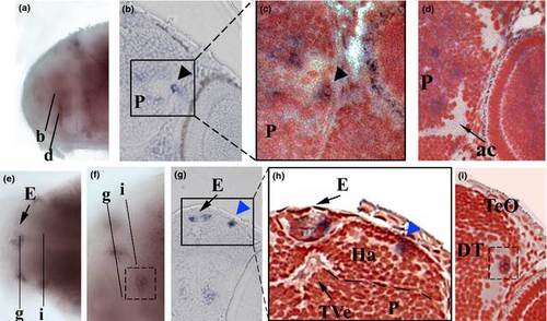
Expression of rxfp3-2b in the anterior region of the 96 h postfertilization (hpf) larval brain. (a) Lateral view of the brain. (b) Transverse section as indicated in (a). (c) Magnification of counter-stained transverse section of the region indicated in (b). (d) Transverse section as indicated in (a). (e) Dorsal view of the larval brain at level of epiphysis. (f) Lateral view of the larval brain at level of epiphysis. (g) Transverse section as indicated in (e) and (f). (h) Magnification of counter-stained transverse section of the region indicated in (g). (i) Particular of counter-stained transverse section as indicated in (e) and (f). Black arrowhead indicates cell cluster probably representing either the telencephalic migrated area or the migrated entopeduncular complex. Blue arrowhead indicates cells in the habenular region. E, epihysis; DT, dorsal thalamus; Ha, habenula; P, pallium; TeO, optic tectum; TVe, telencephalic ventricle. EXPRESSION / LABELING:
|
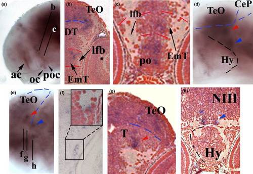
Expression of rxfp3-2b in the central region of the 96 h postfertilization (hpf) larval brain. (a) Lateral view of the brain. (b) Counter-stained transverse section as indicated in (a). (c) Magnification of ventral part of transverse section as indicated in (a). (d) Lateral view of the larval brain at level of hypothalamus. (e) Dorsal view of the larval brain at level of hypothalamus. (f) Transverse section as indicated in (e). In the inset is reported a magnification of counter-stained tranverse section of the region indicated in (f). (g) Counter-stained transverse section as indicated in (e). (h) Counter-stained transverse section as indicated in (e). Blue and red arrowheads indicate cell clusters in the tegmentum region. ac, anterior commissure; CeP, cerebellar plate; DT, dorsal thalamus; EmT, Eminenthia Thalami; Hy, hypothalamus; lfb, lateral forebrain bundle; NIII, nuclei of NIII cranial nerve; oc; optic chiasma; po; preoptic region; poc, postoptic commissure; T, tegmantum; TeO, optic tectum. EXPRESSION / LABELING:
|
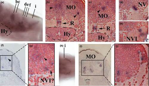
Expression of rxfp3-2b in the posterior region of the 96 h postfertilization (hpf) larval brain. (a) Lateral view of the brain. (b–e) Counter-stained transverse sections as indicated in (a). (f) Transverse section as indicated in (a). (g) Magnification of counter-stained transverse section of the region indicated in (f). (h) Dorsal view of the posterior-most part of the hindbrain. (i) Transverse section as indicated in (h). (j) Magnification of counter-stained transverse section of the region indicated in (i). Black arrowheads indicate cell clusters in the medulla oblongata region. Hy, hypothalamus; MO, medulla oblongata; R, raphe; NV, nuclei of NV cranial nerve; NVI, nuclei of NVI cranial nerve. EXPRESSION / LABELING:
|