
- Title
-
Live imaging of endogenous periodic tryptophan protein 2 gene homologue during zebrafish development
- Authors
- Jayasena, C.S., Trinh, L.A., and Bronner, M.
- Source
- Full text @ Dev. Dyn.

In situ hybridization showing expression comparison between pwp2h in wild-type and citrine expression in the Gt (pwp2h-citrine)ct143a. Lateral and dorsal views are shown for 24 hours post fertilization (hpf) and 48 hpf for pwp2h and citrine. Ventral view at 48 hpf shows citrine expression in the digestive tract. c, cerebellum; e, eye; f, pectoral finbuds; fb, forebrain; i, intestine; is, isthmus: l, liver; m, myotome; p, pancreas; pa, pharyngeal arches; r, retina; t, tectum. Scale bar = 200 μm. EXPRESSION / LABELING:
|
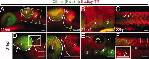
Live confocal imaging of Gt (pwp2h-citrine)ct143a homozygotes. A–F: Bodipy-TR vital dye is used to highlight all cells (red). A,C: Citrine expression at 22 hours post fertilization (hpf), in the head (A) and trunk (C); (B) 24 hpf expression in the pharyngeal arches 1 and 2; (D–F) 31 hpf expression in the head (D,E) and trunk (F). A,D: Projections of confocal z-stacks are shown. B,C,E,F: Projections of confocal z-section. A,D: Pwp2h is expressed at high levels in the tectum compared with the cerebellum. Dotted lines highlight tectum and cerebellum in the embryo. White arrowhead, high expression in the posterior tectum. White arrow, low expression in the anterior tectum. Yellow arrowhead, high expression in the cerebellum. Yellow arrow, drop in Pwp2h expression in the cerebellum. Asterisk: isthmic region. Adjacent image is magnified view of boxed region. D: The magnified view is a confocal z-section. E: Expression in the ear (arrows) and pharyngeal arches 2–7. F: Inset arrowheads: Expression in spinal cord (s) neurons (shown in boxed region). c, cerebellum; m, myotome; n, notochord; ov, otic vesicle; r, retina; t, tectum. Scale bar = 50 μm. EXPRESSION / LABELING:
|

Endogenous Pwp2h in the Gt (pwp2h-citrine)ct143a line at 31 hours post fertilization (hpf) is localized to proliferative domains in the eye (PCNA [proliferating cell nuclear antigen], red). To enhance the Citrine signal, the sections were stained with anti-green fluorescent protein (GFP) antibody (green). All nuclei are stained with DAPI (42,6-diamidino-2-phenylindole; blue). All sections are transverse cryosections. A: Confocal section showing that most of the Citrine tagged Pwp2h (A–A2) is localized to regions also positive for PCNA (A) in the retina (r, highlighted areas). Some expression is also detected in the lens. Inset, high magnification of the boxed region showing nuclei colocalized with PCNA and Citrine. Arrowheads: Citrine localization to areas that appear to correspond to nucleoli. B: Another example through a different axial level of the eye showing colocalization between PCNA and Citrine (highlighted area). Inset: shows Citrine expression only. Scale bars = 50 μm in A,B, 10 μm in inset in A–A2. EXPRESSION / LABELING:
|