
- Title
-
mab21l2 transgenics reveal novel expression patterns of mab21l1 and mab21l2, and conserved promoter regulation without sequence conservation
- Authors
- Cederlund, M.L., Vendrell, V., Morrissey, M.E., Yin, J., Gaora, P.Ó., Smyth, V.A., Higgins, D.G., and Kennedy, B.N.
- Source
- Full text @ Dev. Dyn.
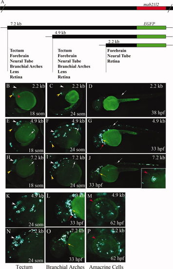
Identification of gene regulatory regions in zebrafish mab21l2. Schematic of the zebrafish mab21l2 gene, and the mab21l2 promoter-reporter constructs and their expression domains (A). Epifluorescent images of wholemount larvae injected with mab21l2:EGFP promoter constructs (B–P). The 2.2-kb construct shows some activity in the forebrain and neural tube (B–D). In contrast, the 4.9- and 7.2-kb constructs direct expression in most known mab21l2expression domains (E–P). At <18–24 somites, they direct EGFP expression in the developing optic tectum, forebrain, and neural tube (E,F,H,I,K,N). At <25 hpf, both promoter constructs drive expression in the lens (inset in J). At <33 hpf, EGFP is still expressed by cells in the optic tectum, branchial arches, neural tube, and in tissue surrounding the heart (G,J,L,O). At <62 hpf, EGFP-positive cells are apparent in the eye (M,P). White arrowheads, tectum; white arrows, neural tube; red arrowheads, eye; red arrows, heart; yellow arrowheads, forebrain; yellow arrows, branchial arches. F,I,K,N, dorsal views; B–D,E,G,H,J,L,M,O,P, lateral views. |

Developmental expression of mab21l1 and mab21l2. At <11 hpf, mab21l2 and mab21l1 are expressed in optic vesicles and tectum (A,B, A2,B2). At <24 hpf, mab21l2, but not mab21l1, is expressed in the lens (C,C2), and only mab21l1 is expressed in olfactory bulbs (D,E, D2,E2). mab21l1 is detected throughout the retina (C2), mab21l2 is confined to the inner retina (C, dots: retina) and both are expressed in tectum and hindbrain (D, D2). At <3 dpf, mab21l1 is expressed in ganglion and amacrine cells (F2), retinal mab21l2 is confined to cells proximal to the lens (F). In the tectum, mab21l1 is widely expressed (G2,H2), whereas only the posterior region expresses mab21l2 (G,H). In the hindbrain, mab21l1 is strongly expressed in the anterior region (G2,H2); mab21l2 is expressed throughout (G2,H2). In branchial arches, mab21l2 is expressed along all craniofacial processes (H); mab21l1 is confined to regions close to the otic vesicle (H2). At 5 dpf, mab21l1 remains expressed in amacrine and ganglion cells, but is downregulated in a ventral patch where mab21l2 is confined (I,I2). mab21l2 expression appears unchanged in the brain but reduced in branchial arches (J,K), mab21l1 has increased in the tectum (J2,K2). Arrowheads (A,B,A2,B2), optic vesicles; arrows (A,B,A2,B2), tectum; arrow (D,E,D2,E2), olfactory bulbs; a, amacrine cells; d, dorsal; g, ganglion cells; v, ventral; tr, temporal retina; nr, nasal retina; l, lens; tm, tectum; hb, hindbrain; ov, otic vesicle; ba, branchial arches; e, eye; cmz, ciliary marginal zone. A,D,G,J,A2,D2,G2,J2: dorsal and B,C,E,F,H,I,K,B2,C2,E2,F2,H2,I2,K2: lateral views. EXPRESSION / LABELING:
|
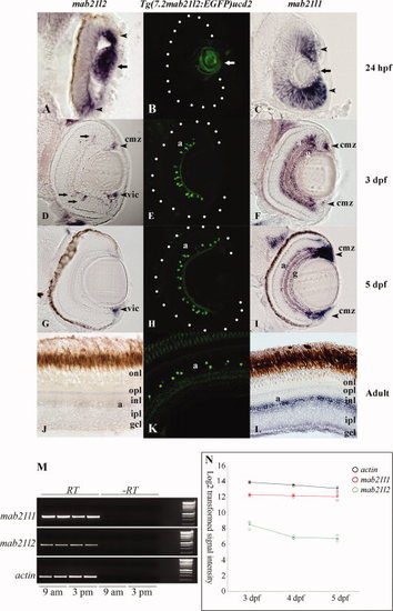
Comparison of the ocular expression of mab21l1, mab21l2, and Tg(7.2mab2ll2:EGFP)ucd2. In <24 hpf eye sections, Tg(7.2mab2ll2:EGFP)ucd2 expresses EGFP in the lens (B), partly recapitulating mab21l2 (A), but not mab21l1, which is widely expressed in the retina (C). At 3 dpf, mab21l2 is weakly expressed in the peripheral INL, dorsal ciliary marginal zone, and ventral iridocorneal canal (D), whereas mab21l1 is upregulated in amacrine and ganglion cell layers (F). From 3 dpf to adult, Tg(7.2mab2ll2:EGFP) is expressed in a subpopulation of amacrine cells in the INL (E,H,K) and thereby partly recapitulates a prominent expression domain of mab21l1 (I,L). In contrast, mab21l2 is only expressed in the ventral iridocorneal canal at <5 dpf (G) and is undetectable in adult sections (J). mab21l1 and mab21l2 expression levels assayed by RT-PCR of 5-dpf eyes. At 5 dpf, mab21l1 is expressed at higher levels than mab21l2. Duplicate RNA samples were isolated at 9 am and 3 pm to confirm that transcription levels did not change during the day. Actin was used as a control (M). mab21l1 and mab21l2 expression levels assayed by microarray analysis of 3–5-dpf eyes This confirmed higher expression levels in the eye of mab21l1 in relation to mab21l2at 3–5 dpf, with decreasing mab21l2 transcript levels after 3 dpf (N). Arrow (A,C), lens; arrowhead (A,C), mab21l1/mab21l2 expression domains; Arrows (D), mab21l2-expressing cells; a, amacrine cell layer; g, ganglion cell layer; cmz, ciliary marginal zone; vic, iridocorneal canal; opl, outer plexiform layer; onl, outer nuclear layer; inl, inner nuclear layer; ipl, inner plexiform layer; gcl, ganglion cell layer. |

mab21l2 is expressed in the ventral iridocorneal canal. A: At 5 dpf, cells expressing mab21l2 localise to the ventral aspect of the eye forming a circumferential structure, distinguished as a section of a channel. Expression within this structure is also detected in posterior serial sections, where it remains at the ventral aspect of the eye contiguous to the iridocorneal angle. B: These sections show a group of cells with a vessel-like shape that can be seen as an ipsilateral section of a tubular structure originating at the iridocorneal angle. The tubular organisation of the cells resembles the ventral canalicular network at the ventral iridocorneal angle, considered homologous to Schlemm′s canal in mammals. |