
- Title
-
A polarised population of dynamic microtubules mediates homeostatic length control in animal cells
- Authors
- Picone, R., Ren, X., Ivanovitch, K.D., Clarke, J.D., McKendry, R.A., and Baum, B.
- Source
- Full text @ PLoS Biol.
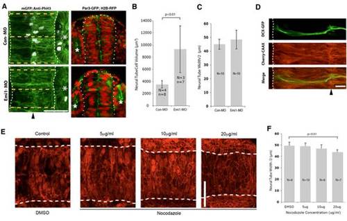
In the left panels neural tubes (outlined with white dots) were visualised with membrane GFP (mGFP), imaged from dorsal view, and measured at three positions (orange dotted lines) around the otic vesicle (asterisk), and the measurements were averaged. Lack of anti-phospho-histone H3 (anti-PhH3) staining in lower panel indicates that Emi1-MO effectively blocks cell divisions. (Midline of neural tube indicated by black arrowhead. Scale bar = 50 μm.) In right panels embryos are expressing Par3-GFP to reveal the nascent apical surface of the neural tube and H2B-RFP to reveal the enlarged nuclei of the division blocked in emi1 morphant embryos (sections viewed in near transverse plane). (B) Cell volume is significantly larger in emi1 morphants (Emi1-MO) than it is in control embryos (Con-MO). (C) Neural tube width was not significantly different between Con-MO and Emi1-MO embryos. (D) Zebrafish embryos were injected with a plasmid expressing DCX-GFP to visualise the microtubule cytoskeleton and mRNA encoding Cherry-CAAX to visualise the neural tube cells. Microtubule bundles were present along the entire length of labelled cells. (Midline indicated by black arrowhead. Scale bar = 10 μm.) (E) Neural tube structure is not affected by treatment with low levels of nocodazole. Zebrafish embryos were injected with mRNA encoding Cherry-CAAX and H2B-RFP and treated with nocodazole at different concentrations. Scale bar: 50 μm. (F) The neural tube of nocodazole-treated embryos was narrower than that of control embryos, and epithelial width was reduced in a concentration-dependent manner. |