
- Title
-
Nasal embryonic LHRH factor plays a role in the developmental migration and projection of gonadotropin-releasing hormone 3 neurons in zebrafish
- Authors
- Palevitch, O., Abraham, E., Borodovsky, N., Levkowitz, G., Zohar, Y., and Gothilf, Y.
- Source
- Full text @ Dev. Dyn.
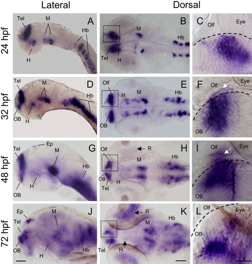
Early developmental expression pattern of the zebrafish nelf by whole-mount in situ hybridization. Subfigures in the right panel are enlargements of the boxed areas in the central panel. Dashed lines depict the forebrain edge. A-C: At 24 hours postfertilization (hpf); D-F: At 32 hpf; G-I: At 48 hpf. nelf is expressed in the olfactory bulb and in the olfactory bulb-olfactory epithelium boundary (white arrows in F and I). J-L: At 72 hpf, nelf is no longer expressed in the olfactory epithelium (L). Ep, epiphysis; H, hypothalamus; Hb, hindbrain; M, midbrain tegmentum; OB, olfactory bulb; Olf, olfactory epithelium; Tel, telencephalon; R, retina. Lateral (left column) and dorsal (central and right panels) views, anterior is left. Scale bar = 100 μM in left and central panels; 50 μM in right panel. EXPRESSION / LABELING:
|

Localization of nelf mRNA (in situ hybridization, red signals) in relation to GnRH3 cells and projections (immunocytochemistry for enhanced green fluorescent protein, green signals) in developing zebrafish. Diagrams show the orientation of the confocal micrographs (dorsal view of embryo head, anterior is top, H-I and K-L are different Z depths). Dashed line depicts the forebrain edge (C,F). A-C: At 26 hpf, GnRH3 cells (arrowheads) are in the olfactory epithelium, nelf mRNA is expressed in the telencephalon. C: Enlargement of boxed area in B. D-F: At 32 hpf, nelf expression in olfactory epithelium around and within GnRH3 neurons (arrowhead), in olfactory bulb (arrows) and presumptive hypothalamus (asterisks). G-I: At 48 hpf, GnRH3 axons project caudally along the path of the terminal nerve and intersect at the optic chiasm (arrowhead) and then proceed to innervate the retina. nelf is expressed within GnRH3 neurons in the terminal nerve (see also Fig. 5D-F). nelf is expressed in the olfactory bulb (arrows), telencephalon domain adjacent to the anterior commissure (asterisks), and in the midbrain (double crossed arrows). J-L: At 72 hpf, GnRH3 neurons in the terminal nerve no longer express nelf. nelf is expressed in olfactory bulb (arrow) and telencephalic domains (asterisks). Scale bar = 20 μM in C,F, 50 μM all other subfigures. EXPRESSION / LABELING:
|

GnRH3-producing neurons transiently express nelf mRNA. A-H: Staining of Tg(gnrh3:EGFP) embryos with fluorescent enhanced green fluorescent protein (EGFP) antibody (A,D,G) and nelf riboprobe (B,E,H). A single optical slice of confocal micrograph (1.8 μm). Dorsal view, anterior to the top. C,F: The analysis reveals colocalization in the olfactory epithelium at 32 hours postfertilization (hpf; C) and in the terminal nerve at 48 hpf (F). At 72 hpf, GnRH3 neurons no longer express nelf (I). Scale bar = 10 μM. |

Representative time-lapse analysis of control-morpholino (MO) and Nelf-MO injected Tg(gnrh3:EGFP) larvae. A-F,H-M: Lateral (A,H) and dorsal (B-F,I-M) views of live larvae head, anterior is left. The injected MO and larvae age are as indicated. A-G: Normal GnRH3 phenotype. A: Bilateral expression of enhanced green fluorescent protein (EGFP) within GnRH3 neurons (white arrows) within the olfactory ephitelium-olfactory bulb boundary. B-F: GnRH3 somata in the telencephalon and projections extend and intersect at the anterior commissure (yellow arrow) and at the optic chiasm (yellow arrowhead). F: The GnRH3 neurons are seen along the GnRH3 axonal pathway and in the hypothalamus (white arrowheads). G: Schematic illustration of GnRH3 cells and projections as shown in F. H-N: Abnormal GnRH3 phenotype. H: Random dispersion of an excess number of GnRH3 neurons in the olfactory area. I-M: GnRH3 projections are absent. The migration of GnRH3 somata is unilateral and does not follow the normal GnRH3 axons outgrowth. The other GnRH3 cells cluster remains at the olfactory organ-olfactory bulb boundary. N: Schematic illustration of GnRH3 cells and projections as shown in M. O: Nelf[Int2Ex3]MO injection altered nelf mRNA splicing as shown by RT-PCR analysis of 1 dpf larvae. P: Sequence analysis indicates that Nelf[Int2Ex3]MO injection interfered with the splicing of exons 2 and 3, causing the addition of 249 nucleotides of intron 2. Upper and lower diagrams represent the cDNA of uninjected and Nelf[Int2Ex3]MO-injected embryos, respectively, as revealed by PCR using specific (arrows). Unfilled rectangles represent exons (lower numbers, bp; upper numbers, exon order), lines represent introns. The oblique lined rectangle represents the transcribed intron 2. EXPRESSION / LABELING:
|