IMAGE

Figure 1

ID
ZDB-IMAGE-201111-1
Source
Figures for Zhang et al., 2020
Image
Figure Caption

Figure 1

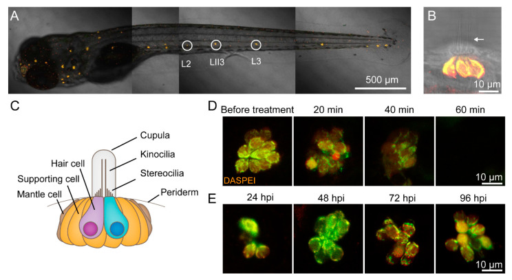
CuSO4 damages hair cells in the lateral line of zebrafish. (A) Lateral line hair cells in a 6 days postfertilization (dpf) wild-type AB zebrafish larva is labeled with 0.05% DASPEI. L2, LII3, and L3 neuromasts are marked with circles. Scale bar represents 500 μm. (B) The lateral view of a neuromast shows sensory hair cells in the center labeled with DASPEI and a bundle of kinocilia (arrow) extending out of the periderm. Scale bar represents 10 μm. (C) A cartoon illustrates the structure of the neuromast. (D) Time lapse imaging shows that when immersed in 5 μM CuSO4 solution, hair cells were gradually injured and damaged within 60 min. Scale bar represents 10 μm. (E) DASPEI staining displays that hair cells regenerate completely within 96 h postinjury (hpi). Scale bar represents 10 μm.

Figure Data
Anatomy Term: neuromast
Acknowledgments
This image is the copyrighted work of the attributed author or publisher, and ZFIN has permission only to display this image to its users. Additional permissions should be obtained from the applicable author or publisher of the image. Full text @ Biomolecules