
Image
|
Figure Caption
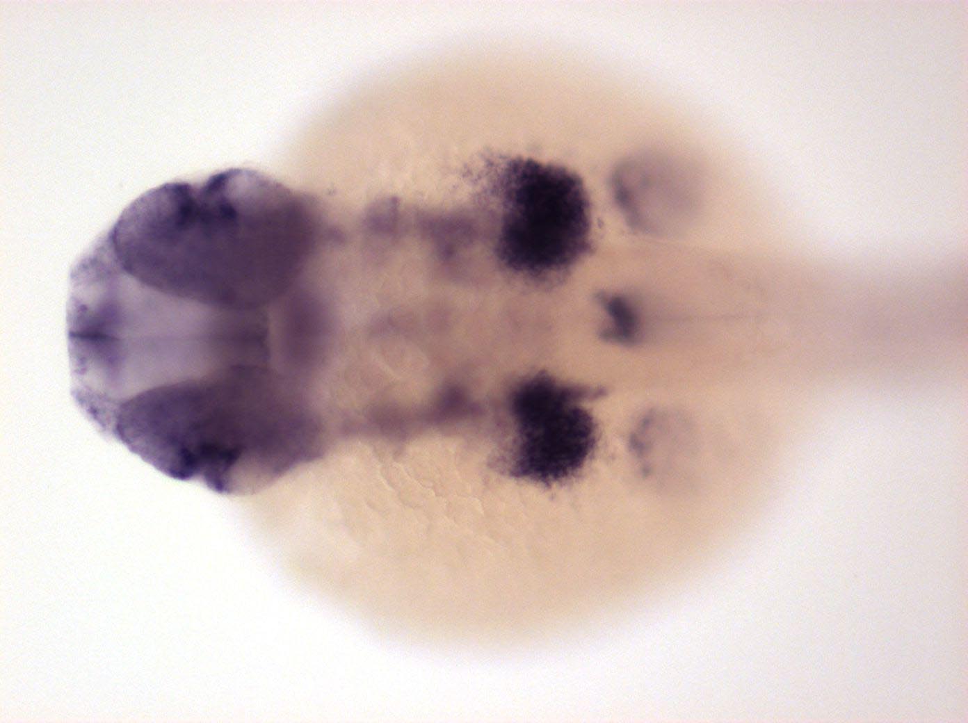
Fig. 5 Expressed in telencephalon, epiphysis, retina, lens, posterior tectum, otic vesicle, rhombomeres boundaries, notochord, pronephric ducts, dorsal spinal cord neurons, mucous cells, pectoral fin bud, one endoderm primordium and very strong in posterior branchial arches
Developmental Stage
Prim-15 to Prim-25
Orientation
| Preparation | Image Form | View | Direction |
| whole-mount | still | dorsal | anterior to left |
Figure Data