
- Title
-
Differential Apoptotic and Mitogenic Effects of Lectins in Zebrafish
- Authors
- Wang, K., Liu, C., Hou, Y., Zhou, H., Wang, X., Mai, K., He, G.
- Source
- Full text @ Front Endocrinol (Lausanne)
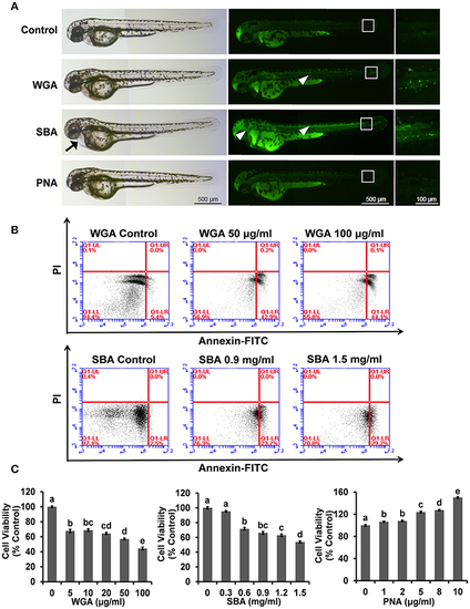
The influence of lectins on the morphogenesis of zebrafish embryos and viability and proliferation in ZFL cells. (A) Zebrafish embryos were treated with indicated lectins at 4 hpf. At 48 hpf, larvae were stained with AO to visualize apoptotic cells which identified as green punctate dots. Arrow indicated the pericardial edema. The boxed region indicated the tail of embryos and the magnification views were shown on the right panel. The arrowhead indicated apoptotic cells in the head and trunk region. Scale bar, 100 and 500 μm. (B) Cells were treated with WGA or SBA for 9 h and stained with Annexin V-FITC/PI for apoptosis analyses using flow cytometry. (C) Cells were treated with series concentrations of WGA or SBA for 9 h. Serum starved cells were treated with indicated concentrations of PNA for 48 h. Cell viability was evaluated by the MTT assay. Results were represented as means with standard errors (n = 6) and analyzed using One-Way ANOVA. Values with different letters in the same column (a–e) were significantly different (p < 0.05) from each other.
|