FIGURE SUMMARY
Title

Fishing for cures: The alLURE of using zebrafish to develop precision oncology therapies

Authors
Astone, M., Dankert, E.N., Alam, S.K., Hoeppner, L.H.
Source
Full text @ NPJ Precis Oncol

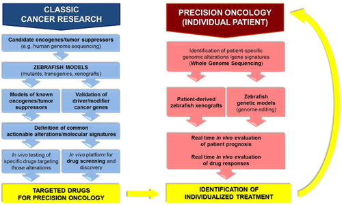
Applications of the zebrafish model in precision oncology. Classic cancer research using zebrafish has contributed to precision oncology through the establishment of numerous cancer models, leading not only to significant advancements in cancer biology, but also to the definition of targeted drugs suitable for personalized cancer treatments (blue, left). Possible applications of zebrafish in the clinic to drive personalized therapies for specific patients have also been shown. The feasibility of this approach has been demonstrated through the use of patient-derived zebrafish xenografts and generation of transgenic zebrafish modeling mutations or translocations defining a specific patient’s tumor (red, right)

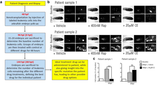
Precision oncology approach to leukemia drug screening using zebrafish. a Flow chart demonstrating the timeline used. Patient-derived leukemia cells were xenotransplanted into zebrafish embryos, which were administered various drugs. Leukemia cell number was used to assess drug efficacy in the zebrafish avatar corresponding to an individual leukemia patient. Assessment of drug efficacy is completed within 8 days, leading to a fast, effective, and individualized cancer treatment. hpf: hours post-fertilization, hpi: hours post-injection of cells, hpt: hours post-treatment. b Bright-field and fluorescence images of zebrafish injected with patient-derived leukemia cells. Embryos were treated with vehicle (control), Rapamycin (Rap) or Compound E (CE). Images were taken at 72 hpt. Scale bars are 500 µM. c A baseline number of leukemia cells was determined at 96 hpi. An increase in the number of leukemia cells when compared to the baseline data demonstrates cell proliferation in the zebrafish model. In patient sample one, data demonstrates a significant (p < 0.0001) response to the Notch inhibition (CE). The patient sample was subsequently sequenced and a gain of function mutation in the Notch pathway was found. Patient sample two did not demonstrate significant results, suggesting the mutation was not in the Notch pathway, which was subsequently confirmed through sequencing. Reproduced with permission and adapted from: Bentley, V.L. et al. Haematologica 100, 70–76 (2015)5

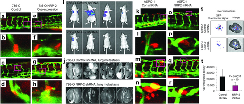
Human cancer cell xenograft models of extravasation in zebrafish and metastasis in mice. ah Human 786-O renal cell carcinoma cells overexpressing retroviral control vector (ad) or neuropilin-2 (NRP-2; eh) were transiently labeled with cell tracker orange dye, microinjected into the pericardium of 3 dpf Tg(Fli-GFP) zebrafish, and imaged 1 day later. ad control 786-O cells stay in the ISVs. e, f 786-O cells overexpressing NRP-2 extravasate from the ISVs. ij 2 × 106 luciferase-labeled 786-O cells suspended in PBS were subcutaneously injected into the right flank of female nude mice. Prior to the tumor growing to 10% of body weight, the subcutaneous tumors were surgically resected. Luciferase imaging was performed on the mice for 4–6 months to monitor metastasis, and the 786-O NRP-2 knockdown group (top) exhibited significantly fewer lung metastases than the control cohort (bottom). kr Human ASPC-1 pancreatic cancer cells were transduced with control shRNA (kn) or NRP-2 shRNA (or), transiently labeled, microinjected, and imaged as described above. k, l Extravasated control shRNA ASPC-1 cells. m, l Actively extravasating control shRNA ASPC-1 cells. or NRP-2 knockdown ASPC-1 cells stay in the ISV. st Male SCID mice were orthotopically injected with 2 × 106 GFP-labeled ASPC-1 pancreatic cancer cells suspended in PBS, and after 15 days liver metastases were assessed by xenogen imaging. Reproduced with permission and adapted from: Cao Y. et al. Cancer Res 73, 4579–4590 (2013)84

Acknowledgments
This image is the copyrighted work of the attributed author or publisher, and ZFIN has permission only to display this image to its users. Additional permissions should be obtained from the applicable author or publisher of the image. Full text @ NPJ Precis Oncol