
- Title
-
Cre inducible site-specific recombination in zebrafish oligodendrocytes
- Authors
- Pinzon-Olejua, A., Welte, C., Chekuru, A., Bosak, V., Brand, M., Hans, S., Stuermer, C.A.
- Source
- Full text @ Dev. Dyn.

mCherry expression in 4- and 6-dpf Tg(mbpa:mCherry-T2A-CreERT2) larvae. A: Scheme of the Tg(mbpa:mCherry-T2A-CreERTT2) construct, which expresses a single open reading frame coding for mCherry (red) and CreERT2 (dark gray) separated by a viral T2A peptide sequence (white) under the control of the Zebrafish mbpa promoter (light gray). B?I: Native mCherry fluorescence in 4-dpf (B?E) and 6-dpf (F?I) transgenic Zebrafish larvae. B,F: Lateral view of the larvae, showing mCherry expression in presumptive myelinating cells. Scale bar, 200?�m. C,D,G,H: Higher magnification of the areas indicated by white boxes in B,F. C,G: mCherry expressing oligodendrocytes in the hindbrain and (D,H) the spinal cord. E,I: Dorsal view of the hindbrain with mCherry fluorescence in presumptive oligodendrocyte cell bodies (arrowhead) and processes (arrow). Asterisks mark auto fluorescence from yolk and pigmentation. Scale bar, 100?�m. |

The mbpa promoter recapitulates expression in mature oligodendrocytes in the larval and adult Zebrafish CNS. A: 6-dpf larvae from the Tg(mbpa:mCherry-T2A-CreERT2) line stained with antibodies against mCherry and MBP show coexistence of mCherry?+?cells (red) in MBP?+?regions (green), suggesting mature oligodendrocytes. Scale bar, 100?�m. B: Higher magnification of the area depicted (white rectangle) in A. Yellow arrowheads mark mCherry-positive oligodendrocytes located in MBP-expressing regions. Scale bar, 50?�m. C: Double-transgenic Tg(mbpa:mCherry-T2A-CreERT2); Tg(mbpa:EGFP) at 7 dpf show co-localization of EGFP and mCherry expression in oligodendrocytes in the spinal cord and hindbrain. D: Higher magnification of the area encircled in C showing EGFP/mCherry-positive oligodendrocytes. E: Scheme of cross-sections of the diencephalon with optic nerve/tract in adult Zebrafish brain as shown in F and G. F: Expression of mCherry in oligodendrocytes of the optic nerve/tract shown by IHC against mCherry (red) and MBP (green) in Tg(mbpa:mCherry-T2A-CreERT2) adult fish. Scale bar, 100?�m. G: Higher magnification of the area depicted (white rectangle) in F showing mCherry-expressing cells (yellow arrowheads) located within the MBP-expressing optic tract. Yellow arrowheads point to oligodendrocytes. Scale bar, 50?�m. H: Cross-section of the retina with the exit point of the optic nerve immunostained for mCherry (red) and MBP (green), showing mCherry-expressing cells within the MBP-expressing processes in the optic nerve as shown in individual panels. Asterisks mark unspecific auto fluorescence of the retinal pigment epithelium, photoreceptors, and extra-retinal tissue. Scale bar, 75?�m. I: Higher magnification of the area depicted (white rectangle) in H showing presumptive oligodendrocytes expressing mCherry (yellow arrowheads) and located within the MBP staining along axons of the optic nerve. DAPI stains all nuclei. Scale bar, 25?�m. |

Conditional CreERT2-mediated recombination in Tg(mbpa:mCherry-T2A-CreERT2) in 5-dpf larvae. A: Schematic representation of treatments applied to achieve conditional CreERT2-mediated recombination with the temperature-inducible, Cre-dependent reporter line Tg(hsp70l:loxP-DsRed-loxP-EGFP). B: Scheme of the ligand-dependent recombination event in double-transgenic Tg(mbpa:mCherry-T2A-CreERT2), Tg(hsp70:loxP-DsRed-stop-loxP-EGFP) larvae. Application of TAM results in Tg(hsp70: loxP-EGFP) in mbpa-expressing cells. C: Lateral view of a 7-dpf larva after 4-OHT treatment and heat shock. Recombination is indicated by a switch from red to green fluorescence in cells in the hindbrain and spinal cord. Scale bar, 200?�m. D,E: Higher magnification of the areas indicated by white boxes in C showing EGFP-expressing oligodendrocytes. F: Dorsal view of the hindbrain with EGFP fluorescence in presumptive oligodendrocyte cell bodies (arrowhead) and processes (arrow). Scale bar, 100?�m. G,H: Recombination can also be observed in Schwann cells myelinating the anterior and posterior lateral lines (arrowheads) Scale bar, 200?�m. I: Recombination only ever occurred in larvae treated with 4-OHT and HS, but never in control siblings treated with ethanol and HS. Scale bar, 200?�m. J: Asterisks mark auto fluorescence from yolk and pigmentation. Scale bar, 200?�m. |
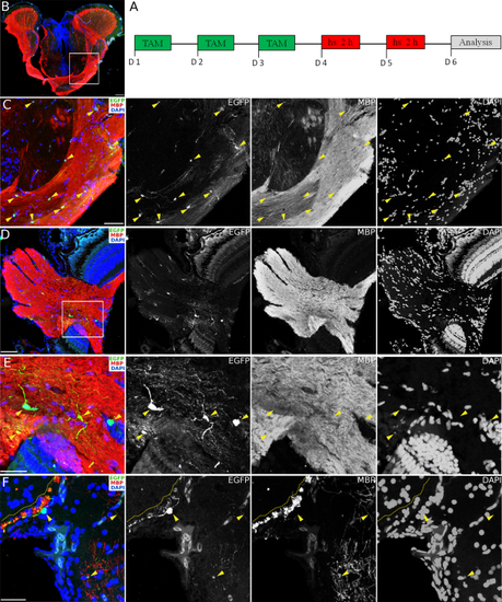
Conditional CreERT2-mediated recombination in Tg(mbpa:mCherry-T2A-CreERT2) in the adult Zebrafish CNS. A: Schematic representation of treatments applied to achieve conditional CreERT2-mediated recombination with the temperature-inducible, Cre-dependent reporter line Tg(hsp70l:loxP-DsRed-loxP-EGFP). B: Cross-section of the diencephalon with optic tracts and the rostral optic tectum immunostained for MBP (red) and EGFP (green). Scale bar, 100?�m. C: Higher magnification of the area depicted (white rectangle) in B showing presumptive oligodendrocytes (marked by arrowheads) after recombination expressing EGFP and co-stained with MBP (red). Scale bar, 50?�m. D: Cross-section of the retina with the exit point of the optic nerve immunostained for EGFP (green) and MBP (red), showing recombined cells (EGFP) within the MBP-expressing optic nerve. Scale bar, 50?�m. E: Higher magnification of the area depicted (white rectangle) in C showing EGFP-expressing cells (yellow arrowheads) co-expressing MBP. Scale bar, 25?�m. F: Cross-section of the dorsal telencephalon showing presumptive mature oligodendrocytes after recombination expressing EGFP (yellow arrowheads), co-immunostained with MBP (red) and located along the MBP-positive lateral olfactory tract (yellow dotted line) and the dorsal part of the entopeduncular nucleus. DAPI stains nuclei. Scale bar, 25?�m. |