
- Title
-
Simple Methods for Generating and Detecting Locus-Specific Mutations Induced with TALENs in the Zebrafish Genome
- Authors
- Dahlem, T.J., Hoshijima, K., Jurynec, M.J., Gunther, D., Starker, C.G., Locke, A.S., Weis, A.M., Voytas, D.F., and Grunwald, D.J.
- Source
- Full text @ PLoS Genet.
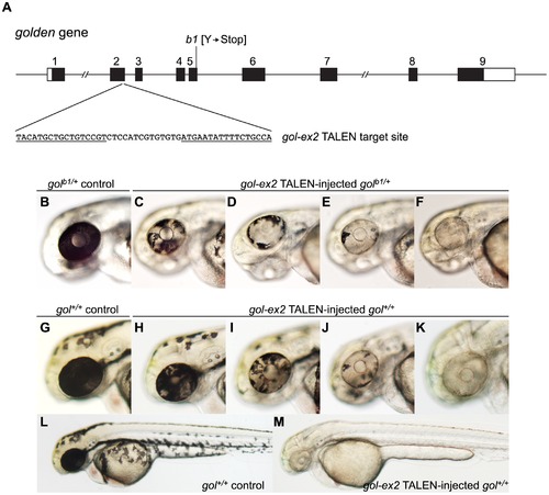
Induction of somatic golden mutations with TALENs. (A) Schematic representation of the genomic structure of the golden (gol) gene, with coding and untranslated exon regions depicted as solid and open boxes, respectively. The locations of the gol-ex2 TALEN target site in exon 2 and the b1 null mutation in exon 5 [33] are indicated. The gol-ex2 TALEN target sequence, with Left and Right TALEN monomer binding sites underlined, is shown. (B–F) Induction of golden mutant cells in the Retinal Pigmented Epithelium (RPE) of heterozygous golb1/+ embryos. Whereas the entire RPE of a 2 dpf heterozygous golb1/+ control embryo (B) is darkly pigmented, the RPEs of golb1/+ embryos injected with gol-ex2 TALEN RNAs (100 pg total) had patches of pigmentless mutant tissue (C–F). (G–M) Induction of golden mutant cells in the soma of wildtype gol+/+ embryos. Wildtype gol+/+ control embryos have darkly pigmented RPEs (G) and dark melanophores scattered over their bodies (L). Following injection at the 1 cell stage with 100 pg gol-ex2 TALEN RNAs, gol+/+ embryos had patches of pigmentless golden mutant tissue in the RPEs (H–K) and some injected embryos appeared entirely devoid of pigmentation (M). PHENOTYPE:
|