
- Title
-
The zebrafish runzel muscular dystrophy is linked to the titin gene
- Authors
- Steffen, L.S., Guyon, J.R., Vogel, E.D., Howell, M.H., Zhou, Y., Weber, G.J., Zon, L.I., and Kunkel, L.M.
- Source
- Full text @ Dev. Biol.
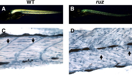
Skeletal muscle is severely disorganized in ruz mutants. (A) Wild-type fish skeletal muscle is highly birefringent at 5–6 dpf. (B) 5–6 dpf ruz homozygous mutants show decreased skeletal muscle birefringence and, on a TL background, body curvature. (C, D) Longitudinal sections of zebrafish skeletal muscle at 5 dpf were stained with hematoxylin. (C) Wild-type muscle shows myofiber alignment between myosepta and peripherally localized nuclei. (D) ruz muscle myofibers appear unaligned with abnormally shaped nuclei. Arrows indicate myosepta. |

Sarcomeric proteins are mislocalized in ruz mutant skeletal muscle. Indirect immunofluorescence was performed on longitudinal sections of 5–6 dpf mutant and wild-type skeletal muscle and samples were imaged at 100× (A, B) or 400x (C–F). (A, C) β-Dystroglycan protein localizes to the myosepta of wild-type embryonic muscle. (B, D) In ruz mutants, β-dystroglycan is maintained at the myosepta. (C) Nebulin localizes to the sarcomere in wild-type embryos. (D) Nebulin sarcomeric localization is lost throughout most of ruz mutant muscle. (E) Skeletal muscle actin similarly localizes to the sarcomere in wild-type embryos while localization is lost in ruz mutants (F). Nuclei were stained with DAPI (blue). |

ruz mutants exhibit decreased expression of specific titin isoforms. (A, B) Indirect immunofluorescence was performed on skeletal muscle longitudinal sections at 6 dpf. Nuclei were stained with DAPI (blue). (A) Wild-type muscle shows titin expression in a repeating sarcomeric pattern. (B) Mutant muscle shows severely decreased titin expression when imaged for the same exposure time. (C) Protein lysates of human psoas muscle (Hu), 2 dpf, 3.5 dpf, and 6.5 dpf wild-type (WT) zebrafish, and 3.5 dpf and 6.5 dpf ruz mutants (ruz) were separated by electrophoresis in adjacent wells of an SDS–agarose gel, transferred to nitrocellulose, and stained with India ink. Lysates loaded either by equal tissue weight per lysis volume or by equal total protein amount showed identical results. At 2 dpf, five faint titin isoforms are apparent. By 3.5 dpf, wild-type fish express four titin isoforms, including two isoforms with the lowest mobility seen during embryonic development. The low mobility doublet is slightly decreased in ruz mutants. At 6.5 dpf, wild-type zebrafish express two titin isoforms (arrows). The larger isoform is severely reduced in ruz mutants while the smaller variant appears increased. Identical results were seen in three independent experiments. |

Morpholinos against TTN1 phenocopy the ruz mutation. (A) 3.5 dpf wild-type fish injected with the TTN1b morpholino against the translational start site of TTN1 (bottom) show decreased birefringence, mobility, and size and are slow to hatch compared with uninjected wild-type siblings (top). Similar results were seen upon injection with the non-overlapping TTN1a morpholino compared with inverted control-injected siblings (data not shown). (B) India ink staining of electrophoretically separated protein lysates of 3.5 dpf TTN1a morphants (MO) and uninjected siblings (uninj) shows loss of the two largest titin isoforms in TTN1 morphants. Smaller titin isoforms are thus likely coded by the adjacent TTN2 gene. (C, D) Longitudinal sections of zebrafish skeletal muscle at 3.5 dpf were stained with hematoxylin. (C) Wild-type muscle shows myofiber alignment between myosepta and peripherally localized nuclei. (D) TTN1a morphants have decreased myofiber alignment and abnormally shaped nuclei similar to ruz mutants. PHENOTYPE:
|
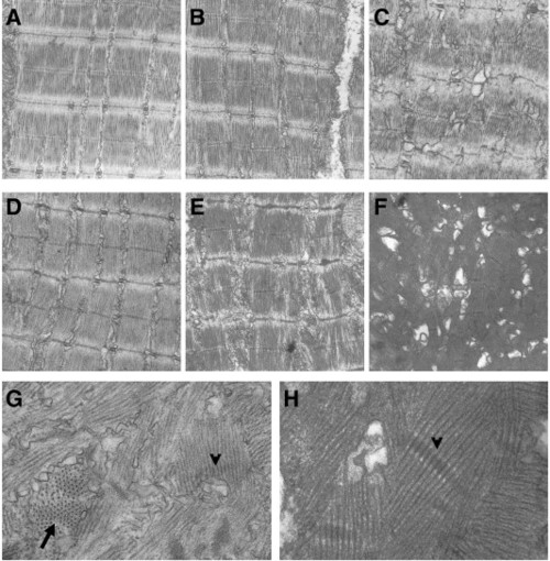
ruz mutants produce normal sarcomeres but show progressive sarcomeric misalignment. (A–H) Longitudinal sections of skeletal muscle from 3.5 dpf (A–C) or 6.5 dpf (D–H) fish were analyzed by transmission electron microscopy. (A) Wild-type fish at 3.5 dpf show normal sarcomeric organization and alignment. (B, C) ruz mutants contain some normal sarcomeres at 3.5 dpf (B) but are also beginning to show sarcomeric disruption (C). At 6.5 dpf, wild-type fish still display normal sarcomeres (D) while ruz mutants contain only rare regions of ordered myofibrils (E). Instead, myofibers contain collapsed sarcomeres with no apparent organization or alignment (F–H). Arrow indicates fibrils perpendicular to the plane of the section. Arrowheads indicate selected Z-disc structures. PHENOTYPE:
|

The ruz dystrophy shows increased calpain-3 and loss of sarcomeric MuRF2 expression. (A) Western blot analysis of 6.5 dpf wild-type and ruz mutants shows a mild increase in calpain-3. Actin expression is used as a loading control. (B, C) Indirect immunofluorescence using MuRF2 antibodies (red) was performed on longitudinally sectioned skeletal muscle of 6.5 dpf fish. Nuclei were stained with DAPI (blue). (B) In wild-type fish, MuRF2 is localized to both the sarcomere (striations) and nucleus (arrow). (C) MuRF2 is lost at the sarcomere in ruz mutants but no significant increase in nuclear localization is detected. PHENOTYPE:
|

Unillustrated author statements PHENOTYPE:
|
Reprinted from Developmental Biology, 309(2), Steffen, L.S., Guyon, J.R., Vogel, E.D., Howell, M.H., Zhou, Y., Weber, G.J., Zon, L.I., and Kunkel, L.M., The zebrafish runzel muscular dystrophy is linked to the titin gene, 180-192, Copyright (2007) with permission from Elsevier. Full text @ Dev. Biol.