ZFIN ID: ZDB-PERS-970109-5
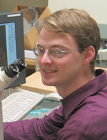
Megason, Sean
Email: megason@hms.harvard.edu
URL: http://www.digitalfish.org/
Affiliation: Megason Lab
Address: Department of Systems Biology Harvard Medical School 200 Longwood Blvd - WAB 536 Boston, MA 02115-5730 USA
Country: United States
Phone: (617) 432-7441
Fax: (617) 432-5012
ORCID ID: 0000-0002-9330-2934


BIOGRAPHY AND RESEARCH INTERESTS
Sean Megason received a B.S. in Molecular Biology from the University of Texas at Austin in 1996 and a Ph.D. in Molecular Cell Biology at Harvard University in 2001. He conducted post-doctoral research at the California Institute of Technology. Sean joined the Department of Systems Biology at Harvard as an Assistant Professor in April 2008.

Research Summary

Embryonic development is the execution of a program encoded in the genome. I wish to understand how this genomic program is executed to create an embryo in the same way that a computer scientist understands the execution of a computer program. The computer scientist has a significant advantage in understanding how a computer program works because the code for a program is transparent: it can be easily read, browsed, altered, and recompiled in its entirety using a software editor. This of course is not the case for biologists because their object of interest, the embryo, is largely a black box. Research in the Megason lab is focused on using two technologies we developed called in toto imaging and flip traps to bring the same level of transparency and quantitation to the code of life. The ultimate goal of in toto imaging is to “upload” the embryo into a digital recreation that can be quantitatively and comprehensively studied with same transparency and facility as a complex computer program.

Our approach to doing systems biology in embryos is heavily based on imaging because of its unique ability to capture quantitative data at single cell resolution in living, functioning embryos. We use zebrafish because of their unique suitability for imaging, genetics, and genomics. We are developing a technology called “in toto imaging” that seeks to track all the cells in a developing tissue and extract quantitative, cell-based data through the use of fluorescent reporters. In toto imaging will allow us to determine complete lineages for organs and to extract cell-based frameworks for use in modeling. We are also developing a technology called FlipTraps. FlipTraps are a novel kind of gene trap with 2 important features: they generate endogenously expressed functional fluorescent fusion proteins, and they generate Cre conditional alleles. We are currently scaling up these efforts as part of the Digital Fish Project which aims to scan in protein expression and mutant phenotype systematically onto a cell-based armature and use these data to construct models that “compute” developmental processes.


PUBLICATIONS
Jia, B.Z., Qi, Y., Wong-Campos, J.D., Megason, S.G., Cohen, A.E. (2023) A bioelectrical phase transition patterns the first vertebrate heartbeats. Nature. 622(7981):149-155
O'Brown, N.M., Patel, N.B., Hartmann, U., Klein, A.M., Gu, C., Megason, S.G. (2023) The secreted neuronal signal spock1 promotes blood-brain barrier development. Developmental Cell. 58(17):1534-1547.e6
Cheng, T., Xing, Y.Y., Liu, C., Li, Y.F., Huang, Y., Liu, X., Zhang, Y.J., Zhao, G.Q., Dong, Y., Fu, X.X., Tian, Y.M., Shu, L.P., Megason, S.G., Xu, P.F. (2023) Nodal coordinates the anterior-posterior patterning of germ layers and induces head formation in zebrafish explants. Cell Reports. 42:112351112351
Wang, A., Zhang, Q., Han, Y., Megason, S., Hormoz, S., Mosaliganti, K.R., Lam, J.C.K., Li, V.O.K. (2022) A novel deep learning-based 3D cell segmentation framework for future image-based disease detection. Scientific Reports. 12:342
Munjal, A., Hannezo, E., Tsai, T.Y., Mitchison, T.J., Megason, S.G. (2021) Extracellular hyaluronate pressure shaped by cellular tethers drives tissue morphogenesis. Cell. 184:6313-6325.e18
Tsai, T.Y., Sikora, M., Xia, P., Colak-Champollion, T., Knaut, H., Heisenberg, C.P., Megason, S.G. (2020) An adhesion code ensures robust pattern formation during tissue morphogenesis. Science (New York, N.Y.). 370:113-116
Mosaliganti, K.R., Swinburne, I.A., Chan, C.U., Obholzer, N.D., Green, A.A., Tanksale, S., Mahadevan, L., Megason, S.G. (2019) Size control of the inner ear via hydraulic feedback. eLIFE. 8:
O'Brown, N.M., Megason, S.G., Gu, C. (2019) Suppression of transcytosis regulates zebrafish blood-brain barrier function. eLIFE. 8:
Ishimatsu, K., Cha, A., Collins, Z.M., Megason, S.G. (2019) Surgical Size Reduction of Zebrafish for the Study of Embryonic Pattern Scaling. Journal of visualized experiments : JoVE. (147):
Swinburne, I.A., Mosaliganti, K.R., Upadhyayula, S., Liu, T.L., Hildebrand, D.G.C., Tsai, T.Y., Chen, A., Al-Obeidi, E., Fass, A.K., Malhotra, S., Engert, F., Lichtman, J.W., Kirchausen, T., Betzig, E., Megason, S.G. (2018) Lamellar projections in the endolymphatic sac act as a relief valve to regulate inner ear pressure. eLIFE. 7
Ishimatsu, K., Hiscock, T.W., Collins, Z.M., Sari, D.W.K., Lischer, K., Richmond, D.L., Bessho, Y., Matsui, T., Megason, S.G. (2018) Size-reduced embryos reveal a gradient scaling based mechanism for zebrafish somite formation. Development (Cambridge, England). 145(11)
Briggs, J.A., Weinreb, C., Wagner, D.E., Megason, S., Peshkin, L., Kirschner, M.W., Klein, A.M. (2018) The dynamics of gene expression in vertebrate embryogenesis at single-cell resolution. Science (New York, N.Y.). 360(6392)
Wagner, D.E., Weinreb, C., Collins, Z.M., Briggs, J.A., Megason, S.G., Klein, A.M. (2018) Single-cell mapping of gene expression landscapes and lineage in the zebrafish embryo. Science (New York, N.Y.). 360(6392):981-987
Hiscock, T.W., Miesfeld, J.B., Mosaliganti, K.R., Link, B.A., Megason, S.G. (2018) Feedback between tissue packing and neurogenesis in the zebrafish neural tube. Development (Cambridge, England). 145(9)
Liu, T.L., Upadhyayula, S., Milkie, D.E., Singh, V., Wang, K., Swinburne, I.A., Mosaliganti, K.R., Collins, Z.M., Hiscock, T.W., Shea, J., Kohrman, A.Q., Medwig, T.N., Dambournet, D., Forster, R., Cunniff, B., Ruan, Y., Yashiro, H., Scholpp, S., Meyerowitz, E.M., Hockemeyer, D., Drubin, D.G., Martin, B.L., Matus, D.Q., Koyama, M., Megason, S.G., Kirchhausen, T., Betzig, E. (2018) Observing the cell in its native state: Imaging subcellular dynamics in multicellular organisms. Science (New York, N.Y.). 360(6386):
Green, A.A., Mosaliganti, K.R., Swinburne, I.A., Obholzer, N.D., Megason, S.G. (2017) Recovery of shape and size in a developing organ pair. Developmental Dynamics : an official publication of the American Association of Anatomists. 246:451-465
Aguet, F., Upadhyayula, S., Gaudin, R., Chou, Y.Y., Cocucci, E., He, K., Chen, B.C., Mosaliganti, K., Pasham, M., Skillern, W., Legant, W.R., Liu, T.L., Findlay, G., Marino, E., Danuser, G., Megason, S., Betzig, E., Kirchhausen, T. (2016) Membrane dynamics of dividing cells imaged by lattice light-sheet microscopy. Molecular biology of the cell. 27(22):3418-3435
Nissim, S., Weeksb, O., Talbot, J.C., Hedgepeth, J.W., Wucherpfennig, J., Schatzman-Bone, S., Swinburne, I., Cortes, M., Alexa, K., Megason, S., North, T.E., Amacher, S.L., Goessling, W. (2016) Iterative use of nuclear receptor Nr5a2 regulates multiple stages of liver and pancreas development. Developmental Biology. 418(1):108-23
Lee, S.G., Huang, M., Obholzer, N.D., Sun, S., Li, W., Petrillo, M., Dai, P., Zhou, Y., Cotanche, D.A., Megason, S.G., Li, H., Chen, Z.Y. (2016) Myc and Fgf Are Required for Zebrafish Neuromast Hair Cell Regeneration. PLoS One. 11:e0157768
Swinburne, I.A., Mosaliganti, K.R., Green, A.A., Megason, S.G. (2015) Improved Long-Term Imaging of Embryos with Genetically Encoded α-Bungarotoxin. PLoS One. 10:e0134005
Xiong, F., Megason, S.G. (2015) Abstracting the principles of development using imaging and modeling. Integrative biology : quantitative biosciences from nano to macro. 7(6):633-42
Xiong, F., Obholzer, N.D., Noche, R.R., Megason, S.G. (2015) Multibow: Digital Spectral Barcodes for Cell Tracing. PLoS One. 10:e0127822
Stooke-Vaughan, G.A., Obholzer, N.D., Baxendale, S., Megason, S.G., Whitfield, T.T. (2015) Otolith tethering in the zebrafish otic vesicle requires Otogelin and α-Tectorin. Development (Cambridge, England). 142(6):1137-45
Hiscock, T.W., Megason, S.G. (2015) Mathematically guided approaches to distinguish models of periodic patterning. Development (Cambridge, England). 142:409-19
Xiong, F., Ma, W., Hiscock, T.W., Mosaliganti, K.R., Tentner, A.R., Brakke, K.A., Rannou, N., Gelas, A., Souhait, L., Swinburne, I.A., Obholzer, N.D., Megason, S.G. (2014) Interplay of Cell Shape and Division Orientation Promotes Robust Morphogenesis of Developing Epithelia. Cell. 159:415-427
Mosaliganti, K.R., Gelas, A., and Megason, S.G. (2013) An efficient, scalable, and adaptable framework for solving generic systems of level-set PDEs. Frontiers in Neuroinformatics. 7:35
Xiong, F., Tentner, A.R., Huang, P., Gelas, A., Mosaliganti, K.R., Souhait, L., Rannou, N., Swinburne, I.A., Obholzer, N.D., Cowgill, P.D., Schier, A.F., and Megason, S.G. (2013) Specified neural progenitors sort to form sharp domains after noisy shh signaling. Cell. 153(3):550-561
Miller, A.C., Obholzer, N.D., Shah, A.N., Megason, S.G., and Moens, C.B. (2013) RNA-seq based mapping and candidate identification of mutations from forward genetic screens. Genome research. 23(4):679-686
Obholzer, N., Swinburne, I.A., Schwab, E., Nechiporuk, A.V., Nicolson, T., and Megason, S.G. (2012) Rapid positional cloning of zebrafish mutations by linkage and homozygosity mapping using whole-genome sequencing. Development (Cambridge, England). 139(22):4280-4290
Xu, C., Fan, Z.P., Müller, P., Fogley, R., Dibiase, A., Trompouki, E., Unternaehrer, J., Xiong, F., Torregroza, I., Evans, T., Megason, S.G., Daley, G.Q., Schier, A.F., Young, R.A., and Zon, L.I. (2012) Nanog-like Regulates Endoderm Formation through the Mxtx2-Nodal Pathway. Developmental Cell. 22(3):625-238
Mosaliganti, K.R., Noche, R.R., Xiong, F., Swinburne, I.A., and Megason, S.G. (2012) ACME: Automated Cell Morphology Extractor for Comprehensive Reconstruction of Cell Membranes. PLoS Computational Biology. 8(12):e1002780
Huang, P., Xiong, F., Megason, S.G., and Schier, A.F. (2012) Attenuation of notch and hedgehog signaling is required for fate specification in the spinal cord. PLoS Genetics. 8(6):e1002762
Trinh, A., Hochgreb, T., Graham, M., Wu, D., Ruf-Zamojski, F., Jayasena, C.S., Saxena, A., Hawk, R., Gonzalez-Serricchio, A., Dixson, A., Chow, E., Gonzales, C., Leung, H.Y., Solomon, I., Bronner-Fraser, M., Megason, S.G., and Fraser, S.E. (2011) A versatile gene trap to visualize and interrogate the function of the vertebrate proteome. Genes & Development. 25(21):2306-20
Agapakis, C.M., Niederholtmeyer, H., Noche, R.R., Lieberman, T.D., Megason, S.G., Way, J.C., and Silver, P.A. (2011) Towards a synthetic chloroplast. PLoS One. 6(4):e18877
Wühr, M., Obholzer, N.D., Megason, S.G., Detrich, H.W., and Mitchison, T.J. (2011) Live imaging of the cytoskeleton in early cleavage-stage zebrafish embryos. Methods in cell biology. 101:1-18
Megason, S.G. (2009) In toto imaging of embryogenesis with confocal time-lapse microscopy. Methods in molecular biology (Clifton, N.J.). 546:317-332
Megason, S.G. and Fraser, S.E. (2003) Digitizing life at the level of the cell: high-performance laser-scanning microscopy and image analysis for in toto imaging of development. Mechanisms of Development. 120(11):1407-1420

NON-ZEBRAFISH PUBLICATIONS
Cedilnik A, Baumes J, Ibanez L, Megason S, Wylie B. (2007). Integration of information and volume visualization for analysis of cell lineage and gene expression during embryogenesis. Proceedings of SPIE Vol. #6809.
Gouaillard A, Brown T, Bronner-Fraser M, Fraser SE, Megason SG. (2007). GoFigure and The Digital Fish Project: Open tools and open data for an imaging based approach to system biology. Insight Journal - special edition “2007 MICCAI Open Science Workshop”. pdf
Megason SG, Fraser SE. (2007). Imaging in Systems Biology. Cell, 130:784-795.
Link BA, Megason SG. (2007) “Zebrafish as a model for development”, in Sourcebook of Models for Biomedical Research, edited by Conn, PM. Humana Press, ISBN: 978-1-58829-933-8.
Megason SG, Amsterdam A, Hopkins N, LinS. (2006). “Uses of GFP in Transgenic Vertebrates” in Green Fluorescent Protein: Properties, Applications and Protocols, Second Edition, Edited by Martin Chalfie and Steven R. Kain. Wiley Press.
Megason SG, Fraser SE. (2003) Digitizing life at the level of the cell: high-performance laser-scanning microscopy and image analysis for in toto imaging of development. Mech Dev. 2003 Nov;120(11):1407-20.
Megason SG, McMahon AP. (2002) A mitogen gradient of dorsal midline Wnts organizes growth in the CNS. Development. 2002 May;129(9):2087-98.