Image
Figure Caption

Figure 6—figure supplement 1. Calcium signaling in individual larvae and neurons.

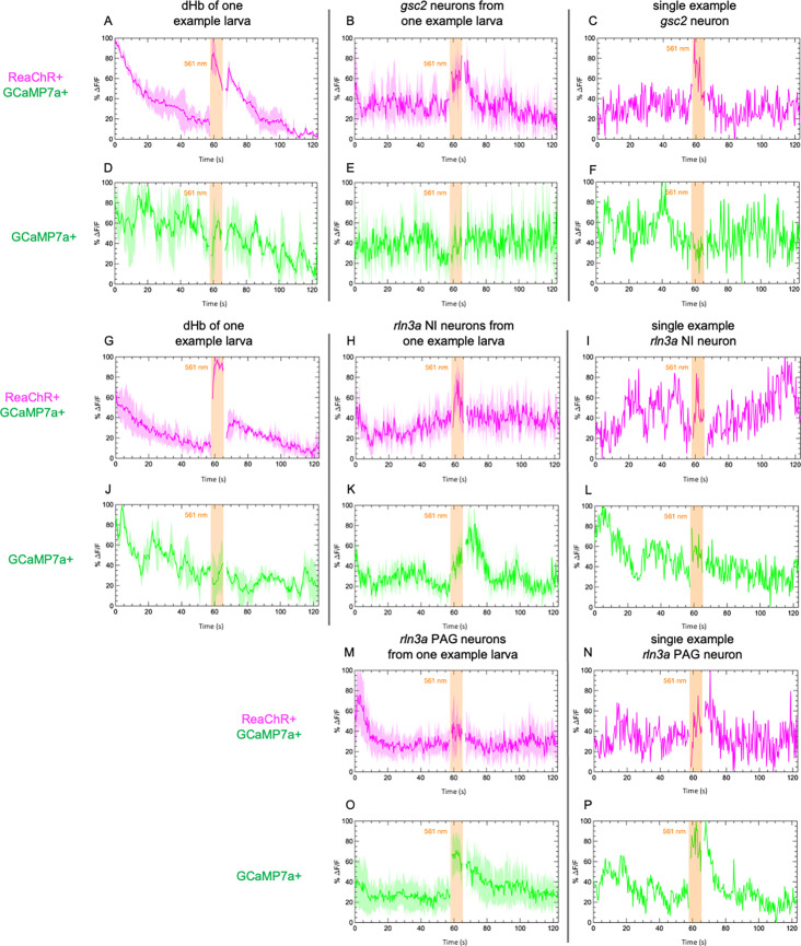
Examples of calcium transients recorded from (A, B, D, E, G, H, J, K, M, O) individual larvae and from (C, F, I, L, N, P) single neurons in additional larvae. Calcium signaling was imaged at 2.6 Hz before, during, and after illumination with 561 nm light in 7 dpf larvae. GCaMP7a signaling (%ΔF/F) is shown for (A, D, G, J) the dorsal habenulae, (B, C, E, F) gsc2 neurons, (H, I, K, L) rln3a NI neurons, and (M, N, O, P) rln3a PAG neurons. Shading indicates standard deviation. Gaps at light onset and offset are due to latency in switching the laser configuration. (A–F) Tg(gsc2:QF2)c721 or (G–P) Tg(rln3a:QF2, he1.1:YFP)c836 driver lines together withTgBAC(gng8:GAL4FF)c426; Tg(UAS:GCaMP7a)zf415; Tg(QUAS:GCaMP7a)c594 with (red trace) or without (green trace) Tg(UAS:ReaChR-RFP)jf50.

Acknowledgments
This image is the copyrighted work of the attributed author or publisher, and ZFIN has permission only to display this image to its users. Additional permissions should be obtained from the applicable author or publisher of the image. Full text @ Elife