
|
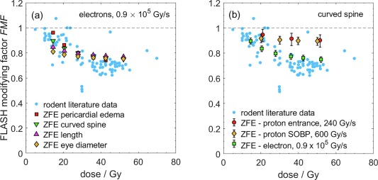
Fig. 3 Panel (a): FLASH modifying factor (FMF) as a function of dose obtained from the dose–effect curves of ZFE irradiated with 30 MeV electrons at UHDR and reference dose rate for four different endpoints: pericardial edema (squares), curved spine (inverted triangles), embryo length deficit (triangles) and eye diameter deficit (diamonds). Panel (b): FMF for the curved spine endpoint as a function of dose for ZFE irradiated with 225 MeV protons at 240 Gy/s (circles), in a proton SOBP at 600 Gy/s (diamonds) and with 30 MeV electrons at 0.9 x 10⁵ Gy/s (squares). The ZFE data is compared with rodent FMF data derived by Böhlen et al. [6] from dose-effect curves found in the literature [1], [32], [39], [40], [41], [42], [43], [44], [45], [46], [47], [48], [49], [50] (light dots). The error bars indicate the uncertainty of the FMF calculated from the dose–effect curves.