
|
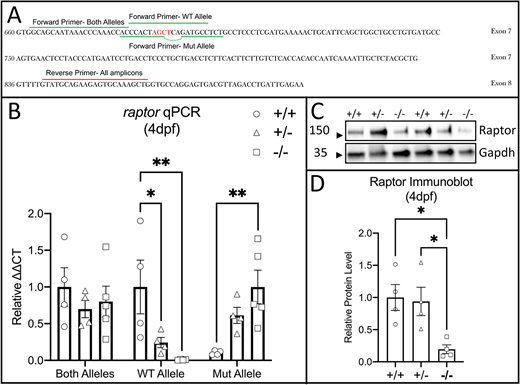
Fig. 2 Validation of the raptor mutant allele. (A) RT-qPCR primer design. Forward primer (green line) amplifying both alleles is upstream of the mutation site. Wild-type allele forward primer (broken green line) contains the nucleotides deleted in the mutant (red); the mutant allele forward primer (green line) matches the deletion. Reverse primer (red line) is in the subsequent exon (exon 8). (B) RT-qPCR using primer pairs that amplify both alleles, the wild-type (WT) allele or the mutant (Mut) allele. cDNA from 4 dpf wild-type fish (circles), 4 dpf heterozygous fish (triangles) or 4 dpf raptor mutant fish (squares) was used. No residual wild-type cDNA is detected in raptor mutants (WT Allele, squares). (C) Immunoblot with a N-terminal anti-Raptor antibody. Total protein lysates were derived from raptor wild types (+/+), heterozygotes (+/−) and mutants (−/−). This blot displays two biological replicates. The membrane was stained using anti-Gapdh antibody as a loading control. The uncropped anti-Raptor blot is shown in Fig. S3. (D) Graph of relative Raptor protein level from the heads of 4 dpf raptor wild-type (circle), heterozygous (triangle) or raptor mutant (square) fish. Raptor protein levels are significantly reduced, albeit not absent, in mutant fish. The statistical analyses were conducted using one-way ANOVA followed by Tukey's honest significant difference (HSD) post-hoc test for multiple comparisons, *P<0.05, **P<0.01. Data are mean±s.e.m.