
|
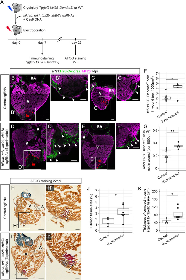
Fig. 7 Loss of hif1ab, nrf1, tbx2b, and zbtb7a affects zebrafish heart regeneration (A) Workflow of adult zebrafish heart injury followed by electroporation of a Cas9 expression vector and a mix of hif1ab, nrf1, tbx2b, and zbtb7a sgRNAs (experimental condition) or a control sgRNA. (B–E) Control (B and C) and experimental (D and E) TgBAC(tcf21:H2B-Dendra2) hearts at 7 dpi, stained against Dendra2 (green) and MF20 (magenta). Shown are hearts with large injuries (B and D) and small injuries (C and E). (B′–E′) show magnifications of the injury areas in (B)–(E), respectively. Arrowheads indicate epicardial cells in the injury area. (F) Quantification of tcf21+ cardiac cell numbers in the injury area (∗p = 0.047). (G) Quantification of tcf21+ cardiac cell numbers in the non-injury area of the ventricle (∗∗p = 0.004). Cell numbers in (F) and (G) have been normalized against injury area (F) and non-injury ventricle area (G). In (F) and (G): n(control) = 7, n(experimental) = 5. (H and I) Acid Fuchsin Orange-G (AFOG) staining showing healthy tissue (orange-brown), fibrin (red), and collagen (blue) in control (H) and experimental (I) hearts at 22 dpi. Asterisks indicate the site of injury. (H′) and (I′) show magnifications of the injury areas in (H) and (I), respectively. Brackets indicate the thickness of the compact muscle layer at the injury site. (J) Quantification of AFOG-stained tissue areas, normalized against ventricle area (∗p = 0.028). (K) Quantification of the thickness of the compact muscle layer at the injury site (∗p = 0.015). In (J) and (K): n(control) = 5, n(experimental) = 7. Scale bars: 100 μm in (B)–(E) and (B′)–(E′), 200 μm in (H) and (I), and 50 μm in (H′) and (I′). A, atrium; BA, bulbus arteriosus; V, ventricle; WT, wild type; ia, injury area. In (F), (G), (J) and (K), box and whiskers plots (in the style of Tukey) indicate median and first/third quartiles. See also Figures S6 and S7 and Table S3.
Reprinted from Developmental Cell, 59(3), Weinberger, M., Simões, F.C., Gungoosingh, T., Sauka-Spengler, T., Riley, P.R., Distinct epicardial gene regulatory programs drive development and regeneration of the zebrafish heart, 351-367.e6, Copyright (2024) with permission from Elsevier. Full text @ Dev. Cell