
|
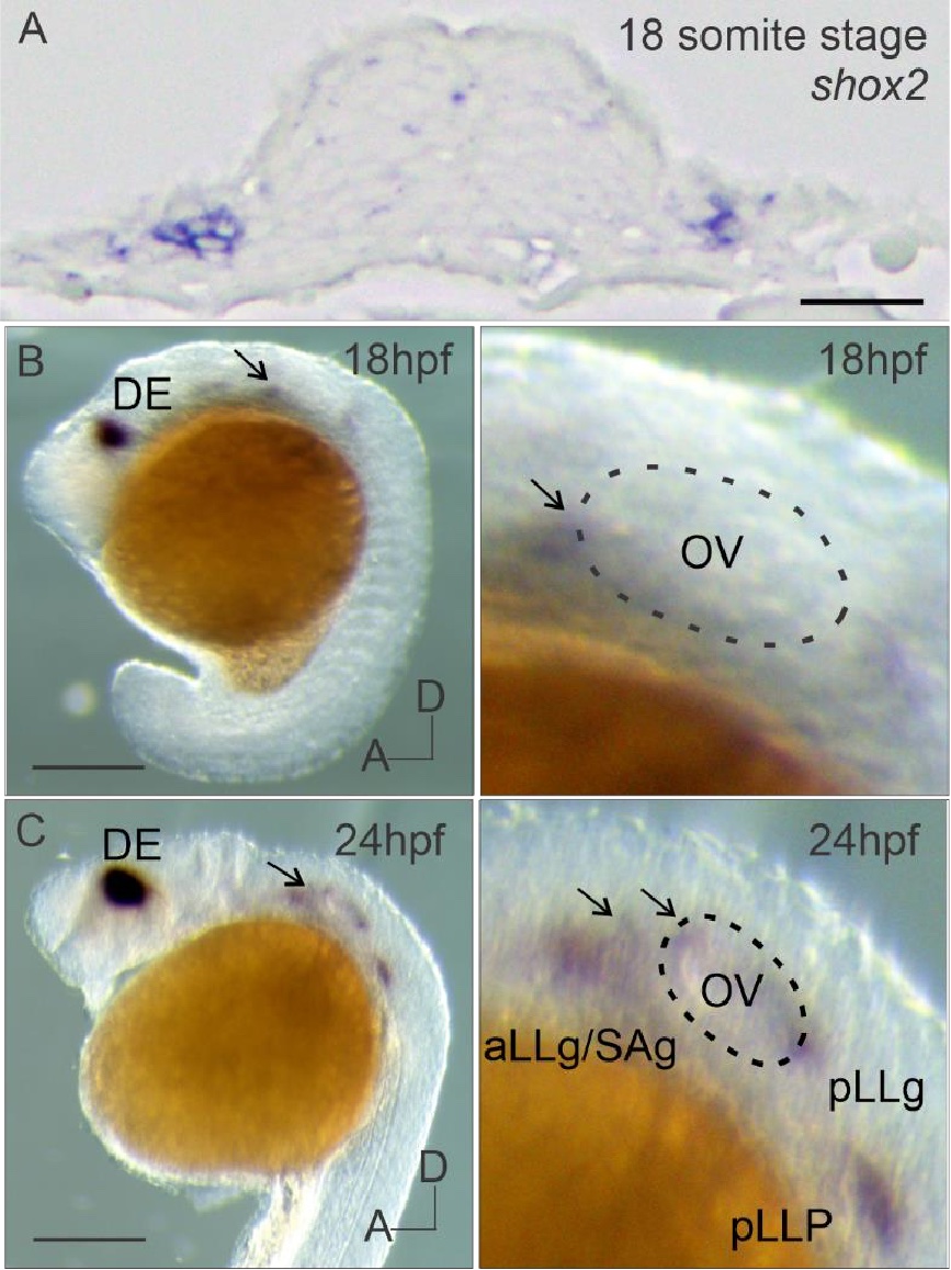
Fig. S1 In situ hybridization of shox2 transcript in the developing zebrafish inner ear (A) shox2 in situ hybridization signal in a section of 18 somite stage (~16 hpf) embryo across the otic placode. Lateral view of whole mount shox2 in situ hybridization signal in zebrafish embryos (left panels) with magnified images of the otic vesicle region (right panels) at (B) 18 and (C) 24 hpf. Arrows point to the anterior ventral region of the otic vesicle. The dashed line in magnified images marks the otic vesicle (18,24 hpf). OV (otic vesicle), DE (diencephalon), aLLG (anterior lateral line ganglia), SAG (statoacoustic ganglion) pLLG (posterior lateral line ganglia) and posterior lateral line primordium (pLLP) are depicted. Anterior (A) and dorsal (D) axis for embryos are labeled. Scale bar: 50 µm.