
Image
|
Figure Caption
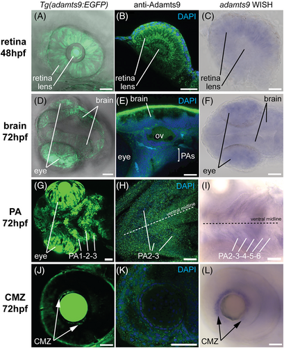
Fig. 4 Comparison of adamts9 signal profile detected by EGFP reporter, immunohistochemistry, and whole mount in situ hybridization (WISH) in retina, brain, pharyngeal arches (PAs), and ciliary marginal zone (CMZ) in zebrafish embryos. A–C, expression in retina. D–F, expression in brain. G–I, expression in PAs. J–L, expression in CMZ. Adamts9 protein was detected using Alexa Fluor 488 conjugated secondary antibody (in green color). Nucleus was co-stained with DAPI. Purple color shows positive WISH signal. CMZ, ciliary marginal zone; OV, otic vesicle; PA, pharyngeal arch. Scale bar: 50 μm.
Acknowledgments
This image is the copyrighted work of the attributed author or publisher, and
ZFIN has permission only to display this image to its users.
Additional permissions should be obtained from the applicable author or publisher of the image.
Full text @ Dev. Dyn.