
Image
|
Figure Caption
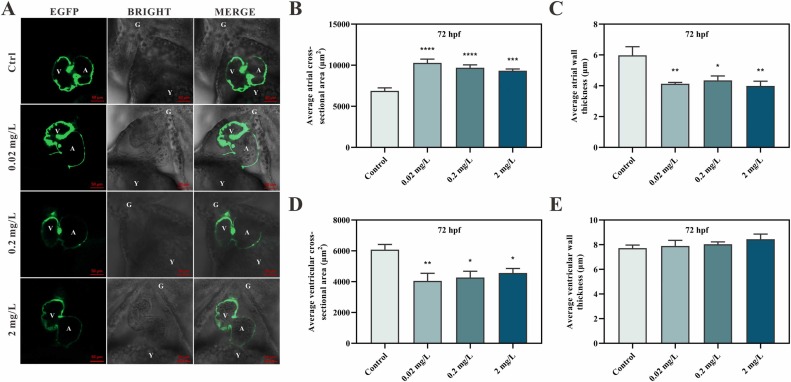
Fig. 3 Cardiac morphogenesis of zebrafish larvae exposed to echimidine for 72 h. (A) Heart morphology of Tg(cmlc2:EGFP) zebrafish exposed to 0.02, 0.2 and 2 mg/L echimidine at 72 hpf. A, atrium; V, ventricle; G, gill; Y, yolk sac. (B) Average atrial cross-sectional area. (C) Average atrial wall thickness. (D) Average ventricular cross-sectional area. (E) Average ventricular wall thickness. The data were analyzed by one-way analysis of variance (ANOVA) followed by Tukey’ s multiple comparisons test and presented as the mean ± SEM (n = 10). * p < 0.05, ** p < 0.01, *** p < 0.001 and **** p < 0.0001.
Acknowledgments
This image is the copyrighted work of the attributed author or publisher, and
ZFIN has permission only to display this image to its users.
Additional permissions should be obtained from the applicable author or publisher of the image.
Full text @ Ecotoxicol. Environ. Saf.