
|
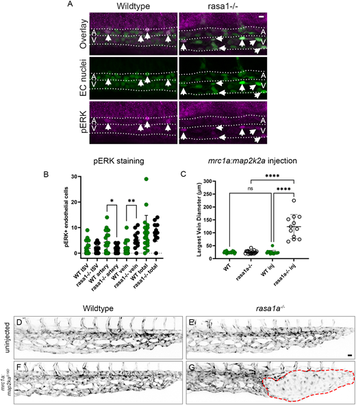
Fig. 7 Ectopic venous activation of pERK in rasa1 mutants drives AVM development. (A) pERK antibody staining in wild type and mutants at 30 hpf. (B) rasa1 mutants show a significant increase in pERK in the vein (wild type, 2.0 cells, rasa1−/−, 5.1 cells; P=0.0020) and a decrease in the DA (wild type, 4.3 cells; rasa1−/−, 1.6 cells; P=0.03) with no change in ISVs at 30 hpf [wild type (n=20), 1.9 cells; rasa1−/− (n=15), 1.4 cells; P=0.5; N=3 experiments, unpaired t-tests]. (C-G) Overexpression of constitutively active map2k2a under a venous promoter (mrc1a) in sensitized rasa1a−/− mutants drives AVM formation. (C) Quantification of largest vein diameter with the ectopic expression of activated mrc1a:map2k2aS219D at 48 hpf. (D,E) Confocal images of uninjected wild type and rasa1a−/− [WTuninj (n=20) versus rasa1a−/−uninj (n=19); P>0.99]. (F,G) Confocal images of wild-type embryos and rasa1a−/− injected with mrc1a:map2k2aS219D [WTinj (n=15) versus rasa1a−/−inj (n=12); P<0.0001; N=2 experiments]. Statistical analysis was carried out using one-way ANOVA with Sidak's correction. Data are mean±s.d. Scale bars: 10 µm in A; 20 µm in D-G.