
|
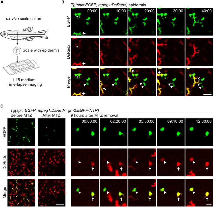
Fig. 4 Mature metaphocytes are generated from spic+ progenitors (A) Schematic outline of the ex vivo scale culture system. Scales with intact epidermis were collected from anesthetized fish and cultured in the L15 medium for time-lapse imaging. (B) Time-lapse imaging of the epidermis of Tg(spic:EGFP;mpeg1:DsRedx) fish. Arrowheads indicate that an EGFP+DsRedx− progenitor cell undergoes cell division to produce two daughter cells, arrows indicate mature metaphocytes with high mobility, and asterisks indicate immature metaphocytes which barely move and have a weak DsRedx signal. Scale bar, 20 μm. (C) Left: representative images of the epidermis of Tg(spic:EGFP;mpeg1:DsRedx;grn2:EGFP-NTR) fish before and after MTZ treatment. Scale bar, 50 μm. Right: time-lapse imaging of the epidermis of Tg(spic:EGFP;mpeg1:DsRedx;grn2:EGFP-NTR) fish starts from 9 h after metaphocytes depletion. The arrowheads indicate an EGFP+DsRedx− progenitor cell that displays and gradually increases the DsRedx signal. The arrows indicate an EGFP+DsRedx+ immature metaphocytes that gradually increases the DsRedx signal. Scale bar, 20 μm.