
|
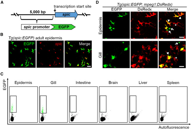
Fig. 3 Tg(spic:EGFP) reporter line specifically marks metaphocytes in the epidermis and gill (A) Schematic outline of the generation of spic reporter line 5 kb promoter sequence immediately upstream of the transcription start site of spic was cloned to drive the expression of EGFP. (B) ISH detection of spic in the epidermis of Tg(spic:EGFP) fish. Scale bar, 20 μm. (C) FACS analysis of spic:EGFP+ cells in adult tissues of the Tg(spic:EGFP) fish. WT ABSR fish without fluorescent signals were utilized as the negative control for gating. (D) Representative images of the epidermis and gill of Tg(spic:EGFP;mpeg1:DsRedx) fish. Asterisks, EGFP−DsRedx+ macrophages; arrowheads, EGFP+DsRedx+ metaphocytes; arrows, EGFP+DsRedx− metaphocyte progenitor cells. Scale bars, 20 μm.