
|
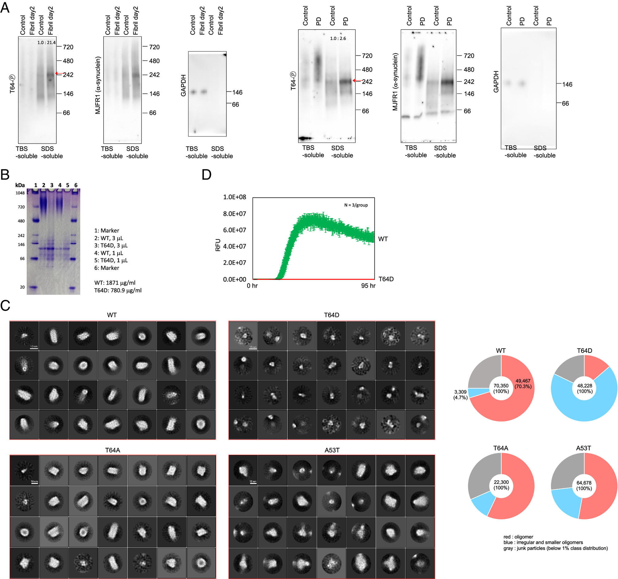
Fig. 3 Oligomer structure of T64-phosphorylated α-synuclein. (A) BN-PAGE and Western blotting of T64-P α-synuclein in the SH-SY5Y cells transfected with α-synuclein fibrils or the human PD brains. α-Synuclein Aggregation Assay Kit was used for the transfection of α-synuclein fibrils and cells are used at 2 d after transfection. TBS-soluble or TBS-insoluble SDS-soluble fractions of the SH-SY5Y cells and human brains were used for BN-PAGE and Western blotting. (B) BN-PAGE and CBB staining analysis of purified α-synuclein oligomers in the WT and T64D phosphomimetic mutant. Each purified α-synuclein (WT or T64D) was incubated for 8 h at 4 °C without agitation and then subjected to ultracentrifugation. The supernatants were subjected to multiple filtration steps to purify α-synuclein oligomers. (C) Two-dimensional electromicroscopic analysis of purified α-synuclein oligomers in the WT, T64A mutant, T64D phosphomimetic mutant and A53T mutant. All particles used for 2D classification were divided into oligomers, particles of lengths of 60 Å or less, and particles with a class distribution of 1% or less, and the number of particles and percentages are plotted in the pie chart. Original electromicroscopic images are shown in SI Appendix, Fig. S5E. (D) Realtime quaking-induced conversion (RT-QuIC) of WT (green) and T64D (red) α-synuclein. RFU: Relative Fluorescence Unit. N = 3 samples/group.