IMAGE

Fig. 7

ID
ZDB-IMAGE-231002-91
Source
Figures for Tsai et al., 2023
Image
Figure Caption

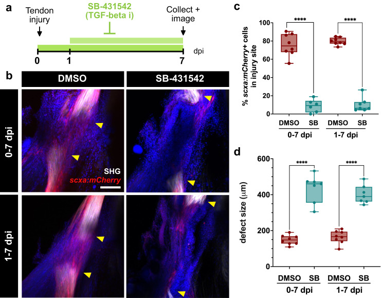
Fig. 7 Canonical TGF-β signaling is required for adult zebrafish tendon regeneration.

a Schematic of experimental design for inhibition of TGF-β signaling during tendon regeneration. b Representative 2-photon stacks of DMSO and SB-431542-treated regenerating tendons at 7 days post-injury (dpi) in scxa:mCherry zebrafish overlaid with second harmonic generation (SHG) signal and Draq5 (in blue) to label nuclei. The 0-7 dpi treatment and 1-7 dpi treatment are shown in the top and bottom panels, respectively. Yellow arrowheads denote the severed tendon ends. Scale bar, 100 µm. c Quantification of the percentage of scxa:mCherry+ cells detected in the injury site at 7 dpi in DMSO and SB-431542-treated zebrafish for both the 0-7 dpi and 1-7 dpi treatments. Unpaired two-tailed t-tests comparing DMSO controls for each treatment with their respective SB-431542-treated counterparts were performed for statistical analysis (0-7 dpi: DMSO, N = 8; SB, N = 6; 1-7 dpi: DMSO, N = 8; SB, N = 7). ****p < 0.0001. d Quantification of the defect size at 7 dpi in DMSO and SB-431542 (SB)-treated zebrafish in both the 0-7 dpi and 1-7 dpi treatments. Unpaired two-tailed t-tests comparing DMSO controls for each treatment with their respective SB-431542-treated counterparts were performed for statistical analysis (0-7 dpi: DMSO, N = 8; SB, N = 7; 1-7 dpi: DMSO, N = 8; SB, N = 7). ****p < 0.0001. Box and whiskers plots in c and d show whiskers extending from the minimum to maximum values, a line at the median, and a box encompassing the 25th to 75th percentiles.

Acknowledgments
This image is the copyrighted work of the attributed author or publisher, and ZFIN has permission only to display this image to its users. Additional permissions should be obtained from the applicable author or publisher of the image. Full text @ NPJ Regen Med