IMAGE

Fig. 5

ID
ZDB-IMAGE-231002-89
Source
Figures for Tsai et al., 2023
Image
Figure Caption

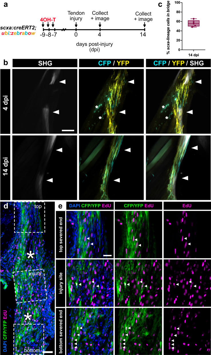
Fig. 5 Pre-existing tenocytes are a major cell source of tendon regeneration.

a Experimental schematic of scxa:creERT2; zebrabow lineage tracing experiment during tendon regeneration. b 2-photon imaging showing CFP+ and/or YFP+ scxa-lineage cells infiltrating the injury site at both 4 and 14 days post-injury (dpi). SHG signal is shown either separately or overlaid. White arrowheads denote severed tendon ends. Asterisks denote autofluorescent blood cells. Scale bar, 100 µm. c Quantification of the percentage of CFP+ and/or YFP+ scxa-lineage cells in the regenerating tendon bridge between the severed tendon ends at 14 dpi (N = 5, mean percentage = 55.88% ± 6.93%). The box and whiskers plot shows whiskers extending from the minimum to maximum values, a line at the median, and a box encompassing the 25th to 75th percentiles. d Representative confocal image of an anti-CFP/YFP stained (in green) section of a regenerating tendon at 4 dpi coupled with EdU labeling (in magenta). Higher magnification of regions of the top severed end, injury site, and bottom severed end are shown in e. Asterisks denote the severed tendon ends. Scale bar, 25 µm. e Higher magnification confocal imaging of CFP/YFP stained cells co-labeled with EdU in the top and bottom severed tendon ends as well as the site of injury. White arrowheads denote examples of CFP/YFP+ cells co-labeled with EdU. Scale bar, 25 µm.

Acknowledgments
This image is the copyrighted work of the attributed author or publisher, and ZFIN has permission only to display this image to its users. Additional permissions should be obtained from the applicable author or publisher of the image. Full text @ NPJ Regen Med